Patient-Specific Disease Modeling with hiPSCs: From Mechanisms to Clinical Translation
Human induced pluripotent stem cells (hiPSCs) have revolutionized biomedical research by enabling the generation of patient-specific cellular models for a wide range of diseases.
Patient-Specific Disease Modeling with hiPSCs: From Mechanisms to Clinical Translation
Abstract
Human induced pluripotent stem cells (hiPSCs) have revolutionized biomedical research by enabling the generation of patient-specific cellular models for a wide range of diseases. This article provides a comprehensive overview of hiPSC technology, from the foundational molecular mechanisms of somatic cell reprogramming to its advanced applications in disease modeling, drug screening, and regenerative medicine. We explore the methodological pipeline for generating and differentiating hiPSCs into various cell lineages, including cardiomyocytes and neurons, and detail their use in modeling neurodegenerative, cardiovascular, metabolic, and autoimmune disorders. The content also addresses key challenges such as genomic instability, immaturity of derived cells, and culture maintenance, while presenting troubleshooting strategies and optimization techniques. Finally, we examine the validation of hiPSC-based models through comparative analyses with primary tissues and discuss their growing role in preclinical drug testing and the development of personalized cell therapies, highlighting recent clinical advances and future directions for the field.
The Foundations of hiPSC Technology: From Reprogramming to Pluripotency
The discovery that somatic cells could be reprogrammed into pluripotent stem cells revolutionized biomedical research, providing an unparalleled tool for patient-specific disease modeling. Human induced pluripotent stem cells (hiPSCs) are somatic cells that have been reprogrammed to an embryonic-like pluripotent state, granting them the capacity to differentiate into any cell type in the body [1] [2]. This technology, pioneered by Shinya Yamanaka and Kazutoshi Takahashi in 2006, effectively bypasses the ethical concerns associated with embryonic stem cells (ESCs) and opens the door to generating patient-specific cell lines for disease research and drug development [3] [2]. Within the context of modern research, hiPSCs provide a critical platform for modeling genetic diseases, screening pharmacological compounds, and developing personalized regenerative therapies, particularly for disorders affecting tissues as complex as the heart and brain [4] [5].
The Discovery and Core Principles of Somatic Cell Reprogramming
Historical Scientific Context
The conceptual foundation for cellular reprogramming was laid decades before the generation of iPSCs. In 1962, Sir John Gurdon demonstrated that the nucleus from a differentiated somatic cell of a tadpole could be transplanted into an enucleated frog egg and generate a whole organism, proving that cellular differentiation is reversible [1]. This principle of somatic cell nuclear transfer was later famously used to clone Dolly the sheep in 1996 [1]. Further evidence emerged in 2001, when Takashi Tada and colleagues showed that fusing adult somatic cells with ESCs could reprogram the somatic nucleus to a pluripotent state [1]. These landmark experiments collectively established that factors within the oocyte and ESC cytoplasm could overwrite the epigenetic landscape of a somatic cell, resetting its developmental clock.
The Yamanaka Breakthrough
Shinya Yamanaka and his team hypothesized that key factors responsible for maintaining pluripotency in ESCs could be sufficient to induce pluripotency in somatic cells. They selected 24 candidate genes with known roles in stem cell identity and maintenance [1]. Using a retroviral vector system to introduce these genes into mouse fibroblasts, they screened for cells that acquired ESC-like properties and could survive antibiotic selection [3] [1]. Through systematic elimination, they identified a core set of four transcription factors that were both necessary and sufficient for reprogramming: Oct3/4, Sox2, Klf4, and c-Myc [3] [1]. The fibroblasts reprogrammed by these "Yamanaka Factors" exhibited the morphology, growth properties, and gene expression markers characteristic of pluripotent stem cells, and could differentiate into tissues of all three germ layers, confirming their pluripotency [3]. For this groundbreaking discovery, Shinya Yamanaka was awarded the Nobel Prize in Physiology or Medicine in 2012, together with Sir John Gurdon [3].
Table 1: The Yamanaka Factors (OSKM)
| Transcription Factor | Full Name | Primary Function in Reprogramming |
|---|---|---|
| Oct3/4 | Octamer-binding transcription factor 3/4 | A POU-homeodomain transcription factor critical for maintaining pluripotency; regulates expression of multiple pluripotency-associated genes. |
| Sox2 | SRY-box transcription factor 2 | A high-mobility-group (HMG) box transcription factor that works synergistically with Oct3/4 to activate pluripotency genes. |
| Klf4 | Kruppel-like factor 4 | A zinc-finger transcription factor that can function as both an activator and repressor; helps initiate the reprogramming cascade. |
| c-Myc | MYC proto-oncogene | A well-known oncogene that enhances proliferation, global histone acetylation, and the efficiency of the reprogramming process. |
Molecular Mechanisms of the Yamanaka Factors
The Yamanaka factors function by orchestrating a profound epigenetic reorganization of the somatic cell genome, silencing genes related to the somatic cell identity while activating the pluripotency network [2]. They achieve this by binding to specific promoter and enhancer regions of target genes.
Oct3/4 and Sox2 form a central core, often acting as a heterodimer to co-occupy and activate the promoters of key pluripotency genes, including their own, creating a positive feedback loop that stabilizes the pluripotent state [3] [2]. Klf4 interacts with this core and can both activate pluripotency genes and repress somatic gene expression. c-Myc, while not strictly essential, dramatically increases reprogramming efficiency by promoting widespread changes in chromatin structure, making gene loci more accessible for transcriptional activation [1]. Together, these factors regulate signaling pathways, microRNAs, and metabolic functions to establish and maintain the self-renewing pluripotent state [2].
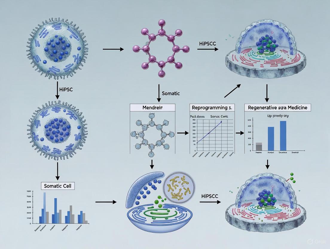
Diagram 1: Somatic cell reprogramming workflow.
Technical Methodologies in hiPSC Generation
Reprogramming Techniques
The initial method of delivering the Yamanaka factors used integrating retroviruses or lentiviruses, which offer high reprogramming efficiency but pose a significant risk of insertional mutagenesis and tumorigenesis due to permanent genomic integration [1] [2]. To address these safety concerns, several non-integrating methods have been developed for clinical applications:
- Episomal Plasmids: Non-integrating DNA vectors that replicate separately from the host genome and are gradually diluted out over cell divisions [2].
- Sendai Virus: An RNA virus that does not integrate into the host genome and is eventually lost from the culture after several passages [2].
- Small Molecules: Chemical compounds that can inhibit DNA methyltransferases or histone deacetylases (e.g., VPA) or modulate signaling pathways (e.g., GSK-3 inhibitor CHIR99021) to enhance reprogramming efficiency and replace one or more reprogramming factors [2].
- Elastin-like Polypeptides (ELP): Used for non-viral transfection to generate virus-free iPSCs [2].
The source of somatic cells can influence the epigenetic memory, heterogeneity, and differentiation potential of the resulting hiPSCs [2]. Common adult cell sources include skin fibroblasts, keratinocytes, and peripheral blood mononuclear cells (PBMCs), chosen for their accessibility and reprogramming efficiency [2]. Modern hiPSC culture often uses feeder-free systems with defined extracellular matrices (e.g., Matrigel, vitronectin) and serum-free media formulations (e.g., Essential 8, mTeSR1) to maintain pluripotency and ensure consistency [2].
Table 2: Comparison of hiPSC Reprogramming Methods
| Method | Mechanism | Advantages | Disadvantages | Typical Efficiency |
|---|---|---|---|---|
| Retro/Lentivirus | Genome-integrating viral vector. | High efficiency; well-established. | Risk of insertional mutagenesis; oncogene reactivation. | High ( ~0.1%) |
| Sendai Virus | Non-integrating RNA virus. | No genomic integration; high efficiency. | Requires effort to clear virus; can be costly. | High |
| Episomal Plasmids | Non-integrating DNA plasmid. | Non-viral; relatively safe. | Lower efficiency compared to viral methods. | Low ( ~0.001%) |
| Small Molecules | Modulates signaling/epigenetics. | Cost-effective; controllable. | Optimization is complex; may not fully replace factors. | Variable |
Characterization and Validation of hiPSCs
Rigorous quality control is essential before hiPSCs can be used for research or therapy. Key characterization steps include:
- Molecular Analysis: Detection and quantification of pluripotency markers (e.g., Oct4, Sox2, Nanog) via immunostaining or PCR [2].
- Functional Assays: Tests like embryoid body formation to confirm the ability to differentiate into cells of all three germ layers [2].
- Karyotyping: Genetic analysis to ensure genomic integrity and absence of major chromosomal abnormalities [6] [2].
hiPSCs in Patient-Specific Disease Modeling and Drug Discovery
The primary application of hiPSC technology is the creation of patient-specific disease models. By reprogramming somatic cells from patients with known genetic mutations, researchers can generate hiPSCs that carry the full genetic background of the disease [4] [5]. These hiPSCs can then be differentiated into relevant cell types—such as cardiomyocytes (hiPSC-CMs) for heart disease or neurons for neurodegenerative disorders—to study disease mechanisms in vitro [4] [7] [5].
Protocol: Disease Modeling with hiPSC-Derived Cardiomyocytes
The following methodology outlines the generation of hiPSC-CMs for disease modeling, incorporating recent advances in suspension culture for improved reproducibility and scale [6].
- hiPSC Quality Control: Begin with quality-controlled master cell banks of hiPSCs. Validate pluripotency marker expression (e.g., >70% SSEA4 positive via FACS) and confirm a normal karyotype. High-quality input cells are critical for successful differentiation [6].
- Formation of Embryoid Bodies (EBs): Dissociate hiPSCs into single cells and culture in stirred suspension bioreactors or spinner flasks to form EBs. Monitor EB diameter, targeting ~100 µm for optimal differentiation efficiency, as smaller EBs may disintegrate and larger EBs suffer from diffusion limits [6].
- Cardiac Differentiation via Wnt Pathway Modulation:
- Mesoderm Induction (Day 0): Add the GSK-3 inhibitor CHIR99021 (e.g., 7 µM) to activate Wnt signaling. This step is critical for directing cells toward the cardiac lineage [6].
- Specification (Day 1-3): After 24 hours, replace medium to remove CHIR99021. Following a 24-hour gap, add the Wnt inhibitor IWR-1 (e.g., 5 µM) for 48 hours to promote cardiac specification [6].
- Maturation and Characterization: Maintain cells in suspension culture with appropriate media. Spontaneous contractions are typically observed around differentiation day 5. Characterize resulting cardiomyocytes by:
This suspension protocol produces hiPSC-CMs with more mature functional properties and lower batch-to-batch variation compared to traditional monolayer differentiation [6].
The Scientist's Toolkit: Essential Reagents for hiPSC-CM Differentiation
Table 3: Key Research Reagents for Cardiac Differentiation
| Reagent / Tool | Function / Application | Example |
|---|---|---|
| Small Molecule GSK-3 Inhibitor | Activates Wnt signaling to induce mesoderm. | CHIR99021 [6] |
| Small Molecule Wnt Inhibitor | Inhibits Wnt signaling to specify cardiac mesoderm. | IWR-1 [6] |
| Stirred Suspension Bioreactor | Enables scalable 3D culture; improves reproducibility and yield. | Commercially available bioreactors or spinner flasks [6] |
| Cardiac Troponin T (TNNT2) Antibody | Validates cardiomyocyte purity and differentiation efficiency via flow cytometry or immunostaining. | Commercial antibodies [6] |
| Pluripotency Marker Antibodies | Quality control of input hiPSCs (e.g., SSEA4). | Commercial antibodies [6] |
Diagram 2: Patient-specific disease modeling workflow.
Current Challenges and Future Directions
Despite significant progress, challenges remain in the field. A primary limitation is the functional immaturity of many hiPSC-derived cell types, including cardiomyocytes, which often resemble fetal rather than adult cells in their structure, metabolism, and electrophysiology [4] [7]. Furthermore, protocol variability and the complexity of reproducing adult disease conditions in a dish can hinder modeling efforts [4]. The future of hiPSC-based disease modeling lies in advancing tissue engineering to create more physiologically relevant 3D models like organoids and engineered heart tissues [4] [7], integrating multi-omics data for a comprehensive view of disease mechanisms [4], and combining hiPSC models with computational tools and AI for high-content phenotypic analysis, such as deep learning-based assessment of sarcomere organization in hiPSC-CMs [8]. As these technologies mature, hiPSCs will continue to be a cornerstone of personalized medicine, enabling deeper understanding of disease pathogenesis and the development of more effective, patient-tailored therapies.
Human induced pluripotent stem cells (hiPSCs) have revolutionized biomedical research by providing a patient-specific platform for disease modeling, drug screening, and regenerative medicine [9] [10]. The reprogramming of somatic cells to a pluripotent state involves profound epigenetic remodeling and dramatic shifts in transcriptional regulation, reverting the cells to a developmentally primitive state [11]. Understanding these molecular mechanisms is paramount for ensuring the fidelity and safety of hiPSCs, particularly for applications in disease modeling where epigenetic abnormalities could compromise experimental validity and clinical potential. This technical guide examines the core epigenetic and transcriptional mechanisms underlying cellular reprogramming, with specific emphasis on implications for generating robust hiPSC models for disease research.
Epigenetic Landscapes in Reprogramming
DNA Methylation Dynamics
The process of reprogramming involves comprehensive reorganization of the DNA methylation landscape. Conventional hiPSCs exist in a "primed" pluripotent state and typically exhibit a hypermethylated genome relative to the somatic cells from which they are derived. Paradoxically, this global hypermethylation occurs alongside a widespread loss of DNA methylation at imprinted loci [11]. Genomic imprinting, a prototypical epigenetic mechanism governing parent-of-origin-specific gene expression, is particularly vulnerable during reprogramming.
More recently, researchers have derived hiPSCs in a more primitive developmental stage termed "naïve pluripotency." These naïve hiPSCs more closely mirror early human embryos and display global genome hypomethylation, which is also accompanied by significant erosion of methylation at imprinted loci [11]. This vulnerability of imprinted control regions is concerning given that loss of imprinting is a well-established feature of several human developmental disorders (such as Beckwith-Wiedemann and Russell-Silver syndromes) and a wide range of cancers [11].
Stability of Imprinted Genes and Associated Pathologies
The stability of imprinting is a critical quality control metric for hiPSCs. Specific imprinted genes commonly misregulated in hiPSCs include those within the DLK1-GTL2 and other imprinted domains, abnormalities which have also been identified in human neoplasms like neuroblastoma, phaeochromocytoma, and Wilms' tumour [11]. The consistent loss of imprinting across different reprogramming methods and pluripotent states indicates a fundamental fragility of these epigenetic marks during the stress of reprogramming.
Table 1: Imprinted Loci Vulnerable During Reprogramming and Their Disease Associations
| Imprinted Locus/Genes | Epigenetic Alteration in hiPSCs | Associated Human Pathologies |
|---|---|---|
| DLK1-GTL2 Domain | Loss of DNA Methylation / Misregulation | Neuroblastoma, Phaeochromocytoma, Wilms' tumour |
| CDKN1C (and other BWS-associated genes) | Loss of DNA Methylation / Allele Imbalance | Beckwith-Wiedemann Syndrome (BWS) |
| Genes in 11p15.5 region | Loss of DNA Methylation | Beckwith-Wiedemann Syndrome, Russell–Silver Syndrome |
Transcriptional Dynamics and Signaling Pathways
The transcriptional reprogramming of a somatic cell to pluripotency is driven by the forced expression of key transcription factors, which activates a cascade of downstream signaling events.
Core Transcriptional Network
The core reprogramming factors—OCT4, SOX2, KLF4, and c-MYC (or alternative combinations)—orchestrate a transcriptional shift by binding to and reactivating the silent pluripotency network. This process involves the silencing of somatic genes and the progressive activation of endogenous pluripotency genes such as NANOG [12]. The successful reactivation of this endogenous network is the hallmark of established hiPSCs.
Signaling Pathways Governing Pluripotency and Differentiation
The signaling environment is critical for both maintaining pluripotency and for the initial steps of differentiation into disease-relevant cell types. Key pathways include:
- TGF-β/ACTIVIN/NODAL Signaling: This pathway is essential for maintaining pluripotency in primed hiPSCs and is a master regulator of germ layer specification. High ACTIVIN A signaling promotes definitive endoderm differentiation, which is the first step toward generating lung, liver, or intestinal organoids [13].
- WNT Signaling: WNT signaling is involved in multiple stages. It is crucial during the initial phase of reprogramming and later, in conjunction with FGF signaling, for patterning the definitive endoderm into anterior and posterior identities [13].
- FGF Signaling: Fibroblast Growth Factor (FGF) signaling works alongside WNT to pattern the endoderm and is also used as a mitogen to promote the proliferation of neural progenitor cells in neural differentiation models [14].
- BMP Signaling: Bone Morphogenetic Protein (BMP) signaling must be inhibited to promote neural ectoderm differentiation, while its modulation is used to target specific sub-populations of mesoderm [13].
The diagram below illustrates the signaling pathways involved in the initial differentiation of hiPSCs into the three germ layers.
Experimental Reprogramming Methodologies
Comparison of Reprogramming Methods
A critical decision in hiPSC generation is the choice of reprogramming method, which can significantly impact genomic integrity and success rates. Early methods using integrating retroviruses or lentiviruses raised concerns about insertional mutagenesis and residual transgene expression [9]. The field has therefore shifted toward non-integrating methods, with Sendai virus (SeV) and episomal vectors being the most prevalent due to their ease of manipulation and relative efficiency [9].
Table 2: Non-Integrating Reprogramming Methods: A Comparative Analysis
| Method | Vector Type | Key Features | Reprogramming Factors Delivered | Reported Success Rate |
|---|---|---|---|---|
| Sendai Virus (SeV) | RNA Virus, Cytoplasmic | Non-integrating, High efficiency, Eventually diluted out by cell division | OCT4, SOX2, KLF4, c-MYC (and EmGFP as reporter) | Significantly higher than episomal method [9] |
| Episomal Vectors | oriP/EBNA-1 Plasmid | Non-integrating, Single transfection possible, Lost at ~5% per cell cycle | OCT4, SOX2, NANOG, LIN28, L-MYC, KLF4, SV40LT [12] | Lower than SeV, but robust [9] |
Detailed Protocol: Episomal Reprogramming of Fibroblasts
The following detailed protocol is adapted from a standard commercial workflow for generating integration-free hiPSCs using episomal vectors [12].
Materials Needed:
- Source Cells: Human fibroblasts (e.g., from skin biopsy).
- Reprogramming Vectors: A mixture of three oriP/EBNA-1 episomal vectors containing the reprogramming genes.
- Culture Media: Fibroblast Medium, Supplemented Fibroblast Medium, N2B27 Medium supplemented with CHALP cocktail, Essential 8 Medium.
- Small Molecules: The CHALP cocktail—CHIR99021 (GSK3β inhibitor), A-83-01 (TGF-β/Activin/Nodal receptor inhibitor), LIF (Leukemia Inhibitory Factor), PD0325901 (MEK inhibitor)—and HA-100 (ROCk inhibitor), and bFGF.
- Transfection System: e.g., Neon Transfection System.
Workflow:
- Day -4 to -2: Plate human fibroblasts to reach 75-90% confluency on the day of transfection.
- Day 0: Harvest and transfect fibroblasts via electroporation with the episomal vector mix. Plate transfected cells onto vitronectin-coated dishes in Supplemented Fibroblast Medium (containing ROCK inhibitor and bFGF).
- Day 1 to 14: Replace medium with N2B27 Medium supplemented with the CHALP cocktail and bFGF. Change medium every other day. The small molecule cocktail greatly improves reprogramming efficiency.
- Day 15: Transition cultures to Essential 8 Medium. Continue feeding every other day while monitoring for the emergence of compact, ESC-like colonies.
- Day ~21 onwards: Manually pick and expand well-defined iPSC colonies onto fresh vitronectin-coated dishes for further characterization and banking.
The overall workflow for generating and differentiating hiPSCs for disease modeling is summarized in the diagram below.
The Scientist's Toolkit: Key Reagents for hiPSC Generation and Differentiation
Table 3: Essential Research Reagents for hiPSC Generation and Early Differentiation
| Reagent Category | Specific Examples | Function in Protocol |
|---|---|---|
| Reprogramming Factors | OCT4, SOX2, KLF4, L-MYC/c-MYC | Master transcription factors that initiate and drive the reprogramming process to pluripotency. |
| Culture Media | Essential 8 Medium, N2B27 Medium | Defined, xeno-free media for robust expansion (E8) and efficient reprogramming/early differentiation (N2B27). |
| Small Molecule Inhibitors/Agonists | CHIR99021 (GSK3βi), PD0325901 (MEKi), A-83-01 (TGF-β Ri), HA-100 (ROCki), SB431542 | Enhance reprogramming efficiency, direct differentiation toward specific germ layers, and improve cell survival. |
| Growth Factors | ACTIVIN A, bFGF, hLIF | Maintain pluripotency (hLIF), promote endoderm differentiation (ACTIVIN A), and stimulate progenitor proliferation (bFGF). |
| Extracellular Matrix | Vitronectin (VTN-N), Geltrex | Provides a defined substrate for the attachment and growth of feeder-free hiPSC cultures. |
Implications for Patient-Specific Disease Modeling
The fidelity of epigenetic reprogramming is directly linked to the utility of hiPSCs in disease modeling. Abnormalities such as loss of genomic imprinting can confound disease phenotypes, especially in models of neurodevelopmental disorders like Autism Spectrum Disorder (ASD) or imprinting disorders themselves [11] [14]. Furthermore, the tendency of hiPSC-derived organoids to resemble fetal rather than adult tissue underscores the importance of ensuring correct epigenetic maturation [13].
In neurodegenerative disease modeling, such as for Parkinson's or Huntington's disease, the goal is to generate authentic neuronal subtypes (e.g., dopaminergic neurons). The differentiation protocols rely on precise transcriptional control and signaling pathway manipulation (e.g., dual SMAD inhibition for neural induction), the efficiency of which can be influenced by the epigenetic state of the starting hiPSCs [10]. Therefore, rigorous quality control, including assessment of genomic imprinting status, DNA methylation patterns, and pluripotency, is essential for generating reliable, reproducible, and clinically predictive hiPSC-based disease models [11] [15].
The field of regenerative medicine has been fundamentally reshaped by the development of technologies that reprogram somatic cells to a pluripotent state. This journey began with somatic cell nuclear transfer (SCNT) and reached a pivotal milestone with the discovery of human induced pluripotent stem cells (hiPSCs). These breakthroughs provided an unprecedented platform for patient-specific disease modeling and circumvented the ethical controversies associated with human embryonic stem cells (hESCs) [16] [17]. The core principle of reprogramming—reverting specialized adult cells to an embryonic-like state—was first demonstrated in pioneering SCNT work, which showed that an intestinal epithelial cell nucleus could be reprogrammed to totipotency when transferred into an enucleated egg, leading to the development of normal tadpoles [17]. This foundational concept laid the groundwork for subsequent technologies, culminating in the landmark discovery that forced expression of specific transcription factors could achieve similar reprogramming without the use of oocytes [17]. This whitepaper traces the critical historical milestones in this field, with a specific focus on their application for developing robust, clinically viable patient-specific disease models.
Foundational Milestone: Somatic Cell Nuclear Transfer (SCNT)
The conceptual and technical foundation for cellular reprogramming was established through SCNT. In this process, the nucleus of a somatic cell is transferred into an enucleated oocyte, which reprograms the somatic nucleus to a totipotent state capable of generating an entire organism [18] [17]. This technology famously led to the creation of Dolly the sheep in 1996, proving that the process was feasible in mammals [17]. While SCNT provided invaluable proof-of-concept for nuclear reprogramming, its application to human therapeutics faced significant hurdles, including technical complexity, limited availability of human oocytes, and substantial ethical concerns [18] [17]. Despite these challenges, SCNT-derived ESCs (SCNT-ESCs) demonstrated functional equivalence to fertilized ESCs, offering potential advantages over early hiPSCs for modeling diseases with strong epigenetic components, as they were thought to avoid the issue of "epigenetic memory" [18].
Table 1: Key SCNT-Derived Pluripotent Stem Cell Features and Implications
| Feature | Technical Implication | Utility for Disease Modeling |
|---|---|---|
| Oocyte-Driven Reprogramming | Complete epigenetic reset | Potentially better modeling of epigenetic diseases |
| Mitochondria from Oocyte | Potential for alloimmunity | Genetic mismatch between donor nucleus and host mitochondria |
| Functional Equivalence to Fertilized ESCs | Gold standard for pluripotency | Faithful developmental model |
| Technical Complexity | Low efficiency; requires specialized expertise | Limited scalability for high-throughput research |
The hiPSC Revolution: From Yamanaka Factors to Clinical-Grade Manufacturing
The Discovery of Induced Pluripotency
A transformative breakthrough occurred in 2006-2007 when Takahashi and Yamanaka demonstrated that the forced expression of four transcription factors—OCT4, SOX2, KLF4, and c-MYC (OSKM)—could reprogram mouse and human somatic fibroblasts into hiPSCs [17]. These cells shared the defining properties of hESCs: self-renewal and pluripotency [19] [16]. This discovery offered a scalable and ethically less contentious path to patient-specific pluripotent cells, immediately positioning hiPSCs as a powerful tool for disease modeling and regenerative medicine [16] [17].
Evolution of Reprogramming Methodologies for Clinical Translation
Early reprogramming methods relied on integrating retroviral vectors, raising safety concerns about insertional mutagenesis and residual transgene expression [19] [20]. The field rapidly advanced to develop non-integrating and footprint-free methods to enhance the safety profile of clinical-grade hiPSCs, as summarized in Table 2.
Table 2: Evolution of hiPSC Reprogramming Methods
| Reprogramming Method | Key Mechanism | Genetic Footprint | Advantages for Clinical/Disease Modeling |
|---|---|---|---|
| Integrating Retrovirus/Lentivirus | Stable genomic integration of OSKM genes | Permanent integration | High efficiency; first reliable method [19] |
| Excisable Vectors (e.g., STEMCCA) | Lentiviral system with LoxP sites for subsequent excision | Transient (removable) | Balances high efficiency with improved safety [20] |
| Non-Integrating Methods (Sendai Virus, Episomal Plasmads) | Viral/plasmid-based transient expression | Footprint-free | No genomic integration; clinically safer [19] [20] |
| Transgene-Free (mRNA, Protein) | Direct delivery of reprogramming mRNAs or proteins | None | Highest safety profile; no foreign genetic material [19] [20] |
The excisable polycistronic stem cell cassette (STEMCCA) represents a critical intermediate technology. This single lentiviral vector expressed the four reprogramming factors and was flanked by LoxP sites, allowing for its subsequent removal via Cre recombinase after reprogramming was complete [20]. This approach significantly reduced the risk of insertional mutagenesis while maintaining high reprogramming efficiency. A key study demonstrated that even when the STEMCCA vector integrated into an intron of an actively transcribed gene (PRPF39), its excision restored the gene's expression to basal levels, generating fully characterized transgene-free hiPSCs that could be differentiated into clinically relevant cell types like cardiomyocytes and neurons [20].
Critical Technical Hurdles and Advanced Solutions
Genetic and Epigenetic Stability
The journey to clinical-grade hiPSCs necessitated a deep understanding of genomic integrity. Studies identified three primary sources of genetic alterations in hiPSCs, as detailed in Table 3.
Table 3: Sources and Mitigation of Genetic Mutations in hiPSCs
| Source of Mutation | Key Findings | Mitigation Strategies |
|---|---|---|
| Pre-existing Somatic Mutations | Mutations in source somatic cells are passively fixed during clonal hiPSC generation [19]. | Use of low-passage, young donor cells (e.g., hematopoietic stem cells) [19]. |
| Reprogramming-Induced Mutations | The reprogramming process itself can generate Copy Number Variations (CNVs) [19]. | Use of non-integrating reprogramming methods [19]. |
| Culture-Acquired Mutations | Extended in vitro passaging can select for advantageous mutations (e.g., in P53) [19]. | Use of low-passage hiPSCs and rigorous genomic monitoring [19]. |
A paramount epigenetic consideration is genomic imprinting, an epigenetic mechanism causing parent-of-origin-specific gene expression. Both conventional ("primed") and more developmentally early ("naïve") hiPSCs frequently show a loss of imprinting, characterized by aberrant DNA methylation at imprinting control regions [19]. Given that loss of imprinting is linked to human developmental disorders and cancers, meticulous monitoring of this epigenetic aberration is essential for the validity of disease models and the safety of future cell therapies [19].
Functional Maturation of hiPSC-Derived lineages
A persistent challenge in using hiPSCs for disease modeling, particularly for late-onset disorders, is the immature phenotype of the differentiated cells. This is strikingly evident in hiPSC-derived cardiomyocytes (hiPSC-CMs), which often resemble fetal rather than adult cardiomyocytes, limiting their efficacy in modeling adult cardiac pathophysiology [21]. Advanced tissue engineering strategies are being deployed to address this. Engineered microenvironments using tunable biomaterials like hydrogels can provide biomechanical cues that drive maturation. For instance, hiPSC-CMs replated on collagen I at later differentiation stages show an upregulation of integrin subunits α1 and β1, activating FAK and ERK signaling pathways—key mechanotransduction cascades that drive structural and electrophysiological maturation [21].
The Scientist's Toolkit: Essential Reagents for hiPSC Research
Table 4: Key Research Reagent Solutions for hiPSC-Based Disease Modeling
| Reagent/Category | Function | Example Application in Disease Modeling |
|---|---|---|
| Reprogramming Factors | Induce pluripotency in somatic cells. | OCT4, SOX2, KLF4, c-MYC (OSKM) are the core factors for generating patient-specific lines [5]. |
| CRISPR-Cas9 System | Genome editing for creating isogenic controls or introducing mutations. | Repairing a disease-causing SNP in a patient hiPSC line to create a genetically matched control [21] [5]. |
| Defined Matrices (e.g., Matrigel, Laminin-521) | Provide a substrate for feeder-free pluripotent cell culture. | Maintaining hiPSCs in a defined, xeno-free culture system suitable for clinical-grade derivation [20]. |
| Lineage-Specific Differentiation Kits | Direct differentiation of hiPSCs toward specific cell fates. | Generating hiPSC-derived cardiomyocytes or neurons for in vitro phenotyping [22] [5]. |
| Multi-Electrode Arrays (MEAs) | Functional electrophysiological analysis of neuronal and cardiac networks. | Detecting aberrant network activity in hiPSC-derived neurons from epilepsy patients [23]. |
Workflow and Signaling in hiPSC Technology
The following diagram illustrates the core experimental workflow for generating and validating patient-specific hiPSCs for disease modeling, integrating the key reagents and quality control steps.
Experimental Workflow for hiPSC Disease Modeling
A critical pathway leveraged in both reprogramming and differentiation is the WNT/β-catenin signaling cascade, which is centrally controlled by integrin-mediated mechanotransduction. The diagram below outlines this key signaling relationship.
Integrin-Mediated Signaling in Fate Control
The path from SCNT to clinical-grade hiPSCs represents a paradigm shift in biomedical research. Current research leverages these patient-specific cells to create increasingly sophisticated in vitro models, particularly for neurological disorders where human tissue is scarce. Innovations include the use of 3D brain organoids and co-culture systems that better mimic the native brain microenvironment [5]. Furthermore, the integration of hiPSC-derived neuronal networks with multi-electrode arrays (MEAs) and advanced computational approaches, such as simulation-based inference, is automating the discovery of disease mechanisms from functional activity data [23] [5]. The convergence of robust clinical-grade manufacturing, precise genome editing, and complex tissue engineering positions hiPSC technology as a cornerstone of modern precision medicine, enabling the deconstruction of disease mechanisms and high-throughput drug screening in a patient-specific context.
The generation of human induced pluripotent stem cells (hiPSCs) has revolutionized biomedical research, enabling patient-specific disease modeling, drug screening, and regenerative medicine approaches. A critical initial decision in hiPSC generation is the selection of the somatic cell source, as this choice impacts reprogramming efficiency, epigenetic memory, and downstream applicability. This technical guide provides an in-depth comparison of three commonly used somatic cell sources: dermal fibroblasts, peripheral blood mononuclear cells (PBMCs), and urinary epithelial cells. Framed within the context of patient-specific disease modeling research, we evaluate these cell sources based on accessibility, reprogramming efficiency, epigenetic characteristics, and suitability for specific disease modeling applications, providing researchers with the necessary information to select the optimal starting material for their experimental needs.
The table below summarizes the key characteristics of the three somatic cell sources, providing researchers with a comprehensive comparison for informed decision-making.
Table 1: Comprehensive Comparison of Somatic Cell Sources for hiPSC Generation
| Parameter | Dermal Fibroblasts | Peripheral Blood Mononuclear Cells (PBMCs) | Urinary Epithelial Cells |
|---|---|---|---|
| Invasiveness of Collection | Moderately invasive (skin punch biopsy) | Minimally invasive (venipuncture) | Non-invasive (voided urine) |
| Primary Cell Types | Fibroblasts | Lymphocytes (T cells, B cells), Monocytes | Podocytes, Proximal Tubular Epithelial Cells (PTECs), other urinary tract epithelia |
| Reprogramming Efficiency | Well-established, reliable | Sendai virus: High efficiency; Episomal: Lower efficiency [9] | Moderate, protocol development ongoing |
| Culture Requirements | Requires expansion in culture prior to reprogramming | Can be reprogrammed directly from fresh or frozen samples [24] | Requires careful culture conditions and expansion [25] |
| Key Markers | Vimentin, Fibroblast Surface Protein | CD45 (pan-leukocyte), CD3 (T cells), CD19 (B cells), CD14 (monocytes) [26] | Podocalyxin, Nephrin (podocytes); AQP1, Megalin (PTECs) [25] |
| Ideal for Disease Modeling | Connective tissue disorders, Fibrotic diseases, General purpose | Hematological disorders, Immunodeficiencies, Autoimmune diseases, Cancer immunology [27] [26] | Genetic kidney diseases, Podocytopathies, Proximal tubulopathies (e.g., Fanconi syndrome) [25] |
| Epigenetic Memory | Tendency towards mesenchymal lineages | Tendency towards hematopoietic lineages [28] | Not well characterized, potentially towards renal/urothelial lineages |
| Primary Challenges | Donor site scarring, Slower expansion | Lower starting material, Complex cell mixture | Lower cell number in urine, Contamination risk, Specialized culture media |
Detailed Methodologies and Workflows
Cell Isolation and Culture Protocols
Dermal Fibroblasts: Fibroblasts are typically isolated from a skin punch biopsy (3-4 mm). The tissue is minced and explants are cultured in fibroblast medium (e.g., DMEM supplemented with 10-15% FBS). Outgrowths of fibroblasts are usually observed within 5-7 days, and cells can be expanded through serial passaging. Fibroblasts are typically used for reprogramming at early passages (P3-P6) to avoid replicative senescence.
PBMCs: PBMCs are isolated from whole blood collected in EDTA or heparin tubes via density gradient centrifugation using media such as HISTOPAQUE-1077 [24]. The blood is diluted and carefully layered over the separation medium, followed by centrifugation. The PBMC layer at the plasma-medium interface is collected, washed, and can be either used fresh or cryopreserved in liquid nitrogen. For reprogramming, PBMCs can be transfected or transduced directly without prior expansion, making the process quicker than with fibroblasts [24] [28].
Urinary Epithelial Cells: Cells are isolated from voided urine samples (typically 50-200 mL). The urine is centrifuged to pellet cells, which are then resuspended and cultured in specialized media. Conditioned media from other cell lines is often used to support growth. A key challenge is the heterogeneous mix of cells in urine, requiring careful characterization and subculture to isolate the desired epithelial populations, particularly podocytes or PTECs for kidney disease modeling [25].
hiPSC Generation via Non-Integrating Methods
Non-integrating reprogramming methods are preferred for clinical applications due to their reduced risk of genomic alterations [9]. The following protocols are applicable to all three somatic cell sources, with variations in initial handling.
Episomal Reprogramming: This method uses OriP/EBNA-1 (Epstein-Barr nuclear antigen-1) based plasmids that deliver reprogramming factors (typically OCT4, SOX2, KLF4, L-MYC, LIN28, and SV40LT) without integrating into the host genome [12]. Cells are transfected via electroporation (e.g., using the Neon Transfection System) and plated onto vitronectin or Matrigel-coated plates. The medium is switched to N2B27 medium supplemented with a cocktail of small molecules (PD0325901, CHIR99021, A-83-01, HA-100, and hLIF) 24 hours post-transfection. After 15 days, the medium is changed to Essential 8 Medium, and emerging iPSC colonies are manually picked around day 21 for expansion and characterization [12].
Sendai Virus (SeV) Reprogramming: This method uses a non-integrating RNA virus to deliver reprogramming factors (OCT4, SOX2, KLF4, and c-MYC). Cells are transduced with the CytoTune Sendai Reprogramming Kit vectors. The medium is refreshed after 24 hours, and cells are cultured for approximately 6 more days with medium changes every other day. The cells are then harvested and replated. Colonies typically appear within 2-3 weeks and are manually picked for expansion [9]. A comparative study found that the Sendai virus method yields significantly higher reprogramming success rates compared to the episomal method [9].
Table 2: Essential Research Reagents for hiPSC Generation
| Reagent Category | Specific Examples | Function in Reprogramming |
|---|---|---|
| Reprogramming Vectors | Episomal iPSC Reprogramming Vectors (Thermo Fisher, A14703), CytoTune Sendai Reprogramming Kit | Delivery of reprogramming factors (OCT4, SOX2, KLF4, c-MYC/L-MYC, LIN28) |
| Cell Culture Media | Essential 8 Medium, N2B27 Medium, Fibroblast Medium (DMEM + 10% FBS) | Support cell survival, proliferation, and create conditions favorable for reprogramming |
| Small Molecule Cocktails | CHALP cocktail (CHIR99021, HA-100, A-83-01, hLIF, PD0325901) | Enhance reprogramming efficiency by modulating key signaling pathways (GSK3β, ROCK, TGF-β, MEK) |
| Culture Substrates | Vitronectin (VTN-N), Geltrex, Matrigel | Provide extracellular matrix support for cell attachment and growth |
| Characterization Reagents | Anti-Tra1-60, Anti-Tra1-81, Anti-SSEA4 antibodies | Confirm pluripotency through immunocytochemistry |
Technical Considerations for Disease Modeling
Source Selection Based on Disease Context: The choice of somatic cell source should be guided by the specific disease being modeled. For hematological and immunological disorders, PBMCs provide a physiologically relevant starting material. Research has shown that PBMCs retain their immune characteristics even in complex diseases; for instance, a study on pituitary neuroendocrine tumors (PitNETs) found distinct transcriptional profiles in patient-derived PBMCs, including upregulated cytokine-receptor interactions [27]. For kidney diseases such as Alport syndrome, podocytopathies, or renal Fanconi syndromes, urine-derived renal epithelial cells offer a direct window into disease pathology [25]. Fibroblasts, while more generic, are valuable for modeling connective tissue disorders and serve as a well-characterized workhorse for general disease modeling.
Addressing Epigenetic Memory: Somatic cells retain an epigenetic memory of their tissue of origin, which can influence the differentiation potential of derived hiPSCs. PBMC-derived hiPSCs may demonstrate a bias toward hematopoietic lineages, while fibroblast-derived hiPSCs may show a propensity for mesenchymal differentiation [28]. This memory can be leveraged to enhance differentiation efficiency toward related lineages but may pose a challenge for targeting unrelated cell types. Extended culture or specific small molecule treatments during reprogramming can help mitigate this bias.
Quality Control and Characterization: Rigorous quality control is essential for all derived hiPSC lines. This includes:
- Pluripotency Marker Confirmation: Immunofluorescence staining for Tra-1-60, Tra-1-81, and SSEA4 [12].
- Genetic Integrity: Karyotyping to ensure chromosomal stability.
- Vector Clearance: PCR analysis to confirm the loss of reprogramming vectors in episomal or Sendai virus methods [9] [12].
- Functional Assessment: Teratoma formation assays or directed differentiation into the three germ layers.
For urine-derived cells specifically, characterization includes quantifying podocytes (using podocalyxin, nephrin) and PTECs (using AQP1, megalin) via qRT-PCR, Western blot, or flow cytometry [25].
Visualizing Workflows and Signaling Pathways
hiPSC Generation Workflow
Key Signaling Pathways in Reprogramming
The selection of an optimal somatic cell source for hiPSC generation is a multifaceted decision that balances practical considerations with specific research goals. Fibroblasts remain a reliable, well-characterized option for general disease modeling; PBMCs offer a minimally invasive source with high reprogramming efficiency, particularly for immunological applications; and urinary epithelial cells provide unique access to renal cell types for kidney disease research without invasive procedures. Understanding the distinct advantages, limitations, and technical requirements of each source enables researchers to design more robust and physiologically relevant patient-specific disease models. As hiPSC technology continues to advance, these somatic cell sources will remain fundamental to unlocking new possibilities in personalized medicine and drug discovery.
hiPSCs in Action: Methodologies and Disease Modeling Applications
The development of human induced pluripotent stem cell (hiPSC) technology has revolutionized biomedical research by providing a patient-specific platform for disease modeling, drug screening, and regenerative medicine [29]. By reprogramming somatic cells into a pluripotent state, researchers can generate virtually any cell type while retaining the complete genetic background of the donor [30] [31]. This capability is particularly valuable for studying human-specific disease mechanisms and developing personalized therapeutic approaches. However, the tremendous potential of hiPSCs depends entirely on the implementation of robust, standardized workflows that ensure the generation of high-quality, fully characterized cells suitable for downstream applications [29]. This technical guide outlines a comprehensive and standardized workflow from somatic cell isolation through reprogramming to directed differentiation, with a specific focus on applications in patient-specific disease modeling research for scientists and drug development professionals.
Strategic Planning and Quality Control
The successful establishment of hiPSC lines requires meticulous strategic planning before initiating experimental work. Researchers should have substantial experience in cell culture techniques and maintain strict adherence to aseptic conditions, especially since hiPSC culture media typically lack antibiotics [29]. A comprehensive quality control framework must be established, including regular karyotyping analyses (recommended at passages 7-10 and every 10-15 subsequent passages) to monitor genomic integrity, short tandem repeat (STR) profiling for cell line authentication, and frequent mycoplasma testing [29]. All equipment and laboratory environments should be properly certified and maintained to ensure consistent cell culture conditions.
Table 1: Critical Quality Control Checkpoints in hiPSC Workflow
| Workflow Stage | QC Checkpoint | Method | Acceptance Criteria |
|---|---|---|---|
| Starting Material | Somatic Cell Viability | Trypan Blue Exclusion | >90% viability |
| Donor Information | Documentation | Complete clinical metadata | |
| Reprogramming | Vector Clearance | RT-PCR [29] | Absence of reprogramming vector |
| Pluripotency Marker Expression | Immunofluorescence/Flow Cytometry [29] | High expression of OCT4, SOX2, NANOG | |
| hiPSC Expansion | Karyotypic Stability | Karyotyping [29] | Normal karyotype |
| In vivo Pluripotency | Teratoma Formation Assay [29] | Differentiation into three germ layers | |
| Differentiation | Lineage Markers | Immunostaining, qPCR | Cell type-specific marker expression |
| Functional Assessment | Cell-type specific assays | Physiological functionality |
Core Methodology: A Standardized hiPSC Workflow
Somatic Cell Isolation and Culture
The hiPSC generation workflow begins with the acquisition of somatic cells from patients or healthy donors. Common sources include peripheral blood mononuclear cells (PBMCs), skin fibroblasts, or keratinocytes. The choice of starting material involves balancing factors such as invasiveness of collection, reprogramming efficiency, and culture requirements. Regardless of source, tissues must be processed using standardized protocols to ensure high cell viability and purity. Isolated somatic cells should be expanded and cryopreserved to create backup stocks before initiating reprogramming.
Reprogramming to Pluripotency
Reprogramming involves the introduction of specific transcription factors (typically OCT4, SOX2, KLF4, and c-MYC) to convert somatic cells to a pluripotent state [29]. Early methods used integrating viral vectors, but current best practices favor non-integrating systems due to safety concerns, including Sendai virus, episomal plasmids, or mRNA transfection [29]. The Sendai viral vector system offers high efficiency and natural clearance through cell divisions, with elimination confirmed via RT-PCR in quality control steps [29]. During reprogramming, emerging hiPSC colonies typically manifest between 14-28 days, with morphological characteristics featuring compact cells with defined borders and high nuclear-to-cytoplasmic ratios.
hiPSC Culture and Maintenance
Maintaining hiPSCs in a pristine, undifferentiated state requires specialized culture conditions. A feeder-free system using Matrigel or similar extracellular matrix components (Geltrex, Vitronectin XF, Laminin-521) with chemically defined media such as Essential 8 (E8) represents the current gold standard [29]. Passaging should employ gentle, non-enzymatic methods like Versene (EDTA solution) to preserve cell viability and prevent spontaneous differentiation [29]. Cultures must be monitored daily, with media changes performed consistently, including weekends. Morphological assessment remains crucial for identifying early differentiation – undifferentiated hiPSCs display compact colonies with clearly defined borders, while differentiated areas appear as loose, non-uniform cells. These differentiated regions should be mechanically removed before passaging.
Table 2: Essential Culture Reagents for hiPSC Maintenance
| Reagent Category | Specific Examples | Function | Technical Notes |
|---|---|---|---|
| Culture Medium | Essential 8 (E8) [29] | Chemically defined medium for maintenance | Supports hiPSC self-renewal without feeders |
| Extracellular Matrix | Matrigel, Geltrex, Laminin-521 [29] | Provides adhesion surface | Mimics basement membrane environment |
| Passaging Reagent | Versene (EDTA) [29] | Gentle cell detachment | Non-enzymatic, improves cell survival |
| Cryopreservation Medium | CryoStor CS10 | Protects cells during freezing | Contains non-penetrating polymers |
| Rho Kinase Inhibitor | Y-27632 [32] | Enhances survival after passaging | Reduces apoptosis in single cells |
Directed Differentiation Strategies
Directed differentiation of hiPSCs leverages developmental principles to guide pluripotent cells through specific lineage pathways using defined cues. The general paradigm involves sequential exposure to small molecules, growth factors, and biophysical cues that mimic embryonic development, typically progressing through intermediate progenitor stages [30].
Cardiac Differentiation
Cardiomyocyte differentiation commonly begins with hiPSCs reaching 80-90% confluence, followed by activation and subsequent inhibition of WNT signaling using small molecules [21] [30]. This precise temporal manipulation drives mesoderm formation, cardiac specification, and ultimately the emergence of spontaneously contracting cardiomyocytes typically within 8-12 days [30]. The resulting hiPSC-derived cardiomyocytes (hiPSC-CMs) demonstrate spontaneous beating and express cardiac markers such as cardiac troponin T and α-actinin, though they predominantly exhibit fetal-like characteristics including small size, disorganized sarcomeres, absent T-tubules, and preferential use of glycolysis over oxidative phosphorylation [30].
Neural Differentiation
Neural differentiation from hiPSCs employs dual SMAD inhibition (using SB431542 and LDN-193189) to suppress transforming growth factor-β and BMP signaling pathways, thereby promoting default neural induction [31] [33]. This approach efficiently generates neural progenitor cells that can subsequently be differentiated into various neuronal subtypes, including cortical neurons, motor neurons, or dopaminergic neurons, through subtype-specific patterning factors [34] [33]. For more complex modeling, 3D cerebral organoids can be generated using techniques that promote self-organization, recapitulating aspects of human brain development and enabling study of cell-cell interactions in a tissue-like context [31].
Advanced Applications in Disease Modeling
Cardiac Disease Modeling
hiPSC technology has profoundly advanced the modeling of inherited cardiovascular diseases such as hypertrophic cardiomyopathy (HCM), dilated cardiomyopathy (DCM), and arrhythmogenic disorders [21] [30]. Patient-specific hiPSC-cardiomyocytes recapitulate key disease phenotypes in vitro, including pathological hypertrophy, contractile dysfunction, and electrophysiological abnormalities [30]. These models are particularly valuable for studying the functional consequences of variants of unknown significance and for patient-specific drug screening and toxicity testing [30]. When combined with genome editing technologies like CRISPR/Cas9 to create isogenic control lines, researchers can definitively establish causal relationships between genetic mutations and disease phenotypes [30].
Neurodevelopmental Disorder Modeling
In neurological research, hiPSC-derived neurons and glial cells have enabled unprecedented study of neurodevelopmental disorders including epilepsy, Tuberous Sclerosis Complex, and intellectual disability disorders [31]. Patient-specific neurons retain the genetic background of the donor, providing direct access to disease mechanisms in human cells [31]. 3D brain organoid models further enhance these studies by recapitulating aspects of cortical development, including progenitor proliferation, neuronal migration, and layer formation, thereby modeling more complex features of neurodevelopmental diseases [31]. The integration of multi-omics approaches with these models helps unravel transcriptional and epigenetic dysregulation underlying disease pathogenesis.
Integration of Tissue Engineering and Maturation Strategies
A significant limitation of conventional 2D hiPSC differentiation is the immature fetal-like state of the resulting cells [30]. Advanced 3D culture systems, including engineered heart tissues, cerebral organoids, and microphysiological systems, enhance cellular maturation by providing more physiological microenvironmental cues [21] [30] [31]. Incorporation of biomechanical stimulation, electrical pacing, and co-culture with non-parenchymal cell types (e.g., cardiac fibroblasts, endothelial cells, or various glial cells) further promotes the development of adult-like functional characteristics [21] [30]. These engineered tissue models more accurately replicate native tissue architecture and function, enabling more clinically predictive disease modeling and drug testing.
The Scientist's Toolkit: Essential Research Reagents
Table 3: Key Reagent Solutions for hiPSC Workflows
| Reagent Type | Specific Examples | Application | Considerations |
|---|---|---|---|
| Reprogramming Vectors | Sendai virus, episomal plasmids, mRNA [29] | Factor delivery for reprogramming | Non-integrating systems preferred for safety |
| Pluripotency Markers | OCT4, SOX2, NANOG, SSEA-4 [29] | Characterization of hiPSCs | Multiple verification methods recommended |
| Cardiac Differentiation | CHIR99021, IWP2, IWR1 [30] | Small molecules for WNT modulation | Precise timing critical for efficiency |
| Neural Differentiation | SB431542, LDN-193189, Noggin [31] | Dual SMAD inhibition for neural induction | High efficiency for neural progenitor generation |
| Extracellular Matrices | Matrigel, Geltrex, Laminin-521 [29] | hiPSC attachment and expansion | Lot-to-lot variability requires testing |
| Cytokines for Hematopoietic/Mesodermal | BMP4, VEGF, FGF2, SCF [32] | Mesoderm and hematopoietic differentiation | Used in specific combinations and sequences |
The standardized workflow from somatic cell isolation to directed differentiation outlined in this technical guide provides a robust framework for generating high-quality hiPSCs and their derivative cell types for disease modeling research. Critical success factors include meticulous attention to quality control, maintenance of optimal culture conditions, and implementation of efficient differentiation protocols. While challenges remain in achieving full maturation of hiPSC-derived cells, ongoing advances in tissue engineering, biomechanical stimulation, and multi-omics integration continue to enhance the physiological relevance of these models. By adhering to standardized methodologies and rigorous characterization standards, researchers can leverage hiPSC technology to advance our understanding of disease mechanisms and accelerate the development of novel therapeutics.
The field of human induced pluripotent stem cell (hiPSC) research has undergone a significant transformation with the advancement of feeder-free and chemically defined culture systems. This evolution is critical for applying hiPSCs to patient-specific disease modeling and regenerative medicine, where reproducibility, scalability, and safety are paramount. Conventional culture systems using feeder cells and serum-containing media present significant challenges for clinical applications due to their undefined nature, batch-to-batch variability, and risk of xenogenic contamination. The development of robust, standardized culture systems eliminates these variables, providing a controlled environment that is essential for rigorous scientific investigation and therapeutic development [35] [36] [37].
This technical guide details the components, protocols, and applications of these advanced culture systems, specifically framed within the context of generating reliable, consistent hiPSC models for disease research and drug development.
Core Components of Advanced hiPSC Culture Systems
Matrices: Replacing Feeder Cells
The transition to feeder-free conditions requires defined matrices to support hiPSC attachment, survival, and self-renewal.
- Recombinant Laminin Fragments: Specifically, laminin-511 E8 fragments (rLN511E8) have been identified as a highly efficient matrix. The E8 fragment is a truncated, recombinantly expressed version that is easier to purify and exhibits superior performance in supporting hiPSC growth as a single-cell suspension. Studies show that hESCs and hiPSCs efficiently form colonies on rLN511E8 but not on other tested matrices like Matrigel or CELLstart. Laminin-511 and -521 interact with α6β1 integrin on hiPSCs, activating the PI3K-Akt pathway to promote survival and growth [35] [38].
- Synthetic Peptide Substrates: Chemically synthesized substrates, such as a vitronectin peptide, provide a fully defined alternative. These peptides support pluripotent cell growth and are compatible with chemically defined media, though some may require optimization for long-term differentiation cultures [38].
Chemically Defined Media
A truly defined system necessitates a media formulation where all components are chemically known.
- StemFit: This xeno-free medium, when used in combination with rLN511E8, supports stable, long-term culture of hiPSCs. Cells can be passaged as single cells for over 20-30 passages while maintaining pluripotency markers and a normal karyotype. The system allows for high expansion rates, with a split ratio of nearly 1:100 and an average doubling time of approximately 28 hours [35].
- CDM3 (Chemically Defined Medium, 3 components): A minimalist medium developed for cardiac differentiation, consisting of only RPMI 1640, L-ascorbic acid 2-phosphate, and rice-derived recombinant human albumin. This formulation, used with small molecule-based differentiation induction, can produce sheets of up to 95% TNNT2+ cardiomyocytes from hiPSCs [38].
- hESF9: A serum-free defined medium used for the generation and maintenance of hiPSCs. It has been successfully employed with non-integrating Sendai virus vectors to reprogram peripheral blood mononuclear cells (PBMCs) on a rLN511E8 substrate [36] [37].
Table 1: Key Components of Feeder-Free and Chemically Defined Culture Systems
| Component Type | Specific Examples | Key Features & Functions | Associated Readouts/Performance |
|---|---|---|---|
| Matrix | Recombinant Laminin-511 E8 (rLN511E8) | Truncated fragment; supports single-cell passaging; binds α6β1 integrin | Colony formation; stable culture >20 passages; split ratio ~1:100 [35] |
| Synthetic Vitronectin Peptide | Fully chemically defined; synthetic | Supports pluripotent growth; may require optimization for long-term differentiation [38] | |
| Chemically Defined Medium | StemFit | Xeno-free; supports self-renewal | Doubling time ~28 hours; maintains pluripotency markers [35] |
| CDM3 | Minimalist (3 components): RPMI 1640, AA 2-P, rHA | Cardiomyocyte differentiation: >85% TNNT2+ cells; yield up to 100 cardiomyocytes per input hiPSC [38] | |
| Reprogramming Vector | Non-integrating Sendai Virus (SeVdp) | Cytoplasmic replication; does not integrate | Generation of hiPSCs from PBMCs and fibroblasts [36] [37] |
Experimental Protocols for hiPSC Generation and Culture
Protocol 1: Generation of hiPSCs from PBMCs under Feeder- and Serum-Free Conditions
This protocol is designed for non-invasive donor sampling and clinical-grade hiPSC generation [36] [37].
- PBMC Isolation and Pre-culture: Isolate PBMCs from whole blood using density gradient centrifugation (e.g., Histopaque 1077). Culture the isolated PBMCs in serum-free medium (e.g., RD6F) supplemented with IL-2 for 6 days.
- Reprogramming: On day 6, transfect 1.0 x 10^5 PBMCs with a non-integrating, replication-defective Sendai virus vector (SeVdp(KOSM)302L) at an MOI of 6. The transfection is performed in a 48-well plate for 2 hours at 32°C.
- Plating and Colony Formation: After transfection, collect the cells by centrifugation and seed them onto a culture vessel coated with recombinant Laminin-511 E8 fragments (0.5 μg/cm²). Use defined medium (e.g., hESF9) and maintain the culture at 38°C. Change the medium every other day.
- Colony Picking and Expansion: Approximately 14 days post-transduction, identify and manually pick hiPSC colonies based on human ESC-like morphology. Mechanically dissociate the picked colonies into small clumps and transfer them to a new Laminin-E8-coated vessel for expansion.
Protocol 2: Maintenance and Passaging of hiPSCs in Feeder-Free Conditions
This protocol describes the long-term culture of established hiPSC lines [35].
- Coating: Coat culture vessels with recombinant Laminin-511 E8 fragments.
- Culture: Maintain hiPSCs in a defined, xeno-free medium such as StemFit.
- Passaging: For passaging, dissociate the hiPSCs into a single-cell suspension using an appropriate dissociation reagent.
- Re-plating: Replete the single cells onto freshly coated vessels at a precise density. A high split ratio of up to 1:100 is achievable due to high cell viability.
- Cryopreservation: Prepare frozen stocks using a standard slow-freezing method. Frozen cells can be thawed directly in a 37°C water bath and recovered in the defined culture system.
Protocol 3: Chemically Defined Cardiac Differentiation
This protocol efficiently generates cardiomyocytes from hiPSCs for disease modeling [38].
- hiPSC Culture: Maintain hiPSCs under chemically defined conditions on a suitable matrix (e.g., rLN521, rLN511).
- Mesoderm Induction (Day 0): Initiate differentiation by adding the GSK3β inhibitor CHIR99021 to CDM3 medium. The optimal seeding density for hiPSCs prior to differentiation is 0.8–1.4 x 10^4 cells/cm².
- Wnt Inhibition (Day 2): After 48 hours of CHIR99021 treatment, add a Wnt production inhibitor (e.g., IWP-2 or Wnt-C59) to the CDM3 medium for 2 days to promote cardiac specification.
- Metabolic Selection (Day 7 onwards): Following the appearance of spontaneous contractions (days 7-9), maintain the cells in CDM3 or a similar defined medium. This can result in cultures of up to 95% TNNT2+ cardiomyocytes, which can be further enriched using metabolic selection methods.
Diagram 1: Cardiac differentiation workflow.
Characterization and Quality Control
Rigorous characterization is essential to confirm the quality of hiPSCs maintained in advanced culture systems.
- Pluripotency Marker Expression: Confirmed via immunocytochemistry for markers such as OCT3/4, NANOG, and TRA-1-60, and by RT-PCR [35] [36].
- In Vitro and In Vivo Differentiation Potential: Assessed via:
- Embryoid Body Formation: Differentiate hiPSCs into cells of all three germ layers (endoderm, mesoderm, ectoderm) and stain for markers like SOX17 (endoderm), α-smooth muscle actin (mesoderm), and βIII-tubulin (ectoderm) [35].
- Teratoma Assay: Inject hiPSCs into immunodeficient mice. Successful teratomas should contain tissues derived from all three germ layers, such as gut-like epithelium (endoderm), cartilage (mesoderm), and neural tissue (ectoderm) [35] [37].
- Karyotype Analysis: Ensure long-term culture does not introduce chromosomal abnormalities. HiPSCs maintained on rLN511E8 with StemFit for over 20 passages have been shown to retain a normal karyotype [35].
- Proteomic Characterization: Global quantitative proteome analysis can be used to develop a protein marker panel for hiPSC characterization, confirming the expression of key pluripotency-associated proteins and ensuring the cells are clearly distinguishable from their somatic cell of origin [39].
Application in Disease Modeling
Defined culture systems are crucial for generating consistent and interpretable data in disease modeling.
- Modeling Inherited Cardiovascular Diseases: HiPSC-derived cardiomyocytes (hiPSC-CMs) generated under defined conditions have been used to model diseases like Hypertrophic Cardiomyopathy (HCM) and Dilated Cardiomyopathy (DCM), recapitulating pathological phenotypes in vitro [4]. The use of a fully defined cardiac differentiation protocol (CDM3) eliminates confounding variables from undefined media components, allowing for clearer insight into disease mechanisms [38].
- Neurological Disease Modeling: HiPSC-derived neural progenitor cells (NPCs) have been used to study schizophrenia. These NPCs, derived and maintained in defined systems, conserved a disease-specific gene signature and exhibited aberrant migration and increased oxidative stress, providing insights into neurodevelopmental mechanisms of the disease [40].
- Muscular Disease Modeling: A simple and efficient method for differentiating hiPSCs into contractile skeletal muscles using inducible MYOD1 expression has been developed. This bulk differentiation system, which minimizes clonal variations, was successfully applied to model Spinal Bulbar Muscular Atrophy (SBMA), demonstrating its utility for pathophysiological analysis [41].
Table 2: Quantitative Outcomes of Advanced Culture and Differentiation Systems
| Process | Input/Scale | Efficiency & Yield | Key Quality Metrics |
|---|---|---|---|
| hiPSC Culture\n(StemFit/rLN511E8) | Single-cell seeding | Split ratio ~1:100; Doubling time ~28.3 hrs [35] | Stable for >20 passages; Normal karyotype; >90% expression of pluripotency markers [35] [39] |
| Cardiac Differentiation\n(CDM3 protocol) | 1.25 x 10^4 hiPSCs/cm² | Yield: up to 1.25 x 10^6 TNNT2+ cells/cm²; Purity: >85-95% TNNT2+ cells [38] | Contractile sheets; Expression of cardiac troponin T; Appropriate response to pathway inhibitors [38] |
| hiPSC Generation from PBMCs\n(SeVdp/Laminin-E8) | 1 x 10^5 PBMCs | Reprogramming efficiency determined by ALP+ colonies [37] | Pluripotency confirmed in vitro and in vivo; Transgene loss confirmed; Normal karyotype [36] [37] |
The Scientist's Toolkit: Essential Research Reagents
Table 3: Key Research Reagent Solutions for Defined hiPSC Culture
| Reagent Category | Specific Product | Critical Function in the Workflow |
|---|---|---|
| Defined Matrix | Recombinant Laminin-511 E8 Fragment | Supports hiPSC adhesion, proliferation, and pluripotency via integrin signaling; enables single-cell passaging. |
| Chemically Defined Medium | StemFit | Provides optimized, xeno-free nutrients and growth factors for robust hiPSC self-renewal. |
| Minimalist Differentiation Medium | CDM3 (RPMI 1640, AA 2-P, rHA) | Serves as a basal medium for highly efficient cardiac differentiation with minimal components. |
| Non-Integrating Reprogramming Vector | Sendai Virus (SeVdp) Vector | Delivers reprogramming factors (OCT4, SOX2, KLF4, c-MYC) without genomic integration for safe hiPSC generation. |
| Small Molecule Inducers | CHIR99021 (GSK3β inhibitor) | Activates Wnt signaling to robustly induce mesoderm at the start of cardiac differentiation. |
| Myogenic Differentiation Factor | Doxycycline-inducible MYOD1 | Master regulator that directly transdifferentiates hiPSCs into the skeletal muscle lineage. |
Signaling Pathways in Defined Differentiation
Understanding the signaling pathways manipulated during differentiation is key to protocol optimization. The cardiac differentiation protocol is a prime example of targeted pathway manipulation.
Diagram 2: Signaling pathways in cardiac differentiation.
The diagram illustrates the critical pathways:
- Biphasic Wnt Manipulation: The protocol hinges on the precise temporal control of canonical Wnt signaling. Initial activation using a GSK3β inhibitor like CHIR99021 is essential for mesoderm induction. Subsequent inhibition of Wnt production (e.g., with IWP-2) is required for cardiac specification from the mesoderm [38].
- Essential Supporting Pathways: Inhibition studies during the mesoderm induction phase (days 0-2) have demonstrated that FGF, BMP, and Activin/NODAL signaling are also essential for efficient cardiac differentiation, likely part of a paracrine feedback loop initiated by Wnt activation [38].
The adoption of feeder-free conditions and chemically defined media is no longer an aspiration but a necessary standard for rigorous, reproducible, and clinically relevant hiPSC-based research. These systems provide the foundation for reliable patient-specific disease modeling, accurate drug screening, and the future development of cellular therapeutics. By eliminating the variability and unknown factors associated with feeders and serum, researchers can have greater confidence that the phenotypes observed in their hiPSC-derived models are a true reflection of the underlying genetics and disease pathology, rather than an artifact of the culture environment.
The study of neurodegenerative diseases has been transformed by the advent of human induced pluripotent stem cell (hiPSC) technology, which enables the generation of patient-specific neural cells for disease modeling and drug discovery. Traditional approaches relying on postmortem human tissue or animal models have significant limitations; postmortem tissue only reveals end-stage pathology, while animal models fail to capture the full spectrum of human disease due to critical species-specific differences [42]. hiPSCs, generated by reprogramming human somatic cells into a pluripotent state, can be differentiated into various neural cell types, including neurons, astrocytes, and microglia, providing unprecedented opportunities to study human neurodegenerative diseases in vitro [42] [43]. This technical guide examines the application of hiPSC-based models to three major neurodegenerative disorders: Alzheimer's disease (AD), Parkinson's disease (PD), and amyotrophic lateral sclerosis (ALS), with a focus on advanced methodologies, disease-specific phenotypes, and their applications in therapeutic development.
Alzheimer's Disease Modeling
Pathophysiology and hiPSC Applications
Alzheimer's disease is characterized by progressive memory loss and cognitive decline, with pathological hallmarks including amyloid-β (Aβ) plaques, neurofibrillary tangles composed of hyperphosphorylated tau, and neuroinflammation. Genome-wide association studies (GWAS) have identified numerous AD risk genes, many of which are highly expressed in glial cells, particularly astrocytes [44]. Major AD risk genes such as APOE, CLU, and FERMT2 are predominantly expressed in astrocytes, highlighting their critical role in disease pathogenesis [44] [45].
hiPSC-based models have revealed that astrocytes contribute significantly to AD pathology through multiple mechanisms. Healthy hiPSC-derived astrocytes play important roles in APP processing and secrete Aβ, contributing to its accumulation in AD brains [44] [45]. Astrocytes derived from early-onset AD patients carrying PSEN1 mutations display AD hallmarks including increased Aβ and reactive oxygen species (ROS) production, altered inflammatory responses, and dysregulated calcium homeostasis compared to isogenic controls [44]. When co-cultured with neurons, PSEN1 mutant astrocytes induce alterations in neuronal calcium signaling, demonstrating their profound impact on neuronal physiology [44].
APOE4 Astrocyte Dysfunction
The APOE ε4 allele represents the strongest genetic risk factor for late-onset AD, and hiPSC studies have elucidated its specific effects on astrocyte function. hiPSC-derived astrocytes carrying the APOE4 variant produce and secrete less APOE protein compared to APOE3 astrocytes, are less efficient at clearing extracellular Aβ, and show impaired lipid/cholesterol metabolism [44] [45]. APOE4 astrocytes also exhibit increased inflammatory responses associated with Transgelin 3 (TAGLN3) downregulation and NF-κB activation [44]. This proinflammatory state can be pharmacologically reverted by TAGLN3 supplementation, identifying it as a potential therapeutic target [44]. In co-culture systems, APOE4 astrocytes provide less support for neuronal survival and synaptogenesis while exacerbating neuroinflammation [44] [45]. Cerebral organoids containing APOE4 neurons and astrocytes show increased synapse loss, neurodegeneration, and tau pathology compared to APOE3 organoids [44].
Multi-Cellular Interactions and Advanced Modeling
The complexity of AD pathogenesis necessitates models that capture cell-cell interactions. In tri-culture systems with hiPSC-derived neurons, astrocytes, and microglia, the complement protein C3—elevated in AD brains and involved in neurodegeneration—increases under inflammatory conditions due to astrocyte-microglia reciprocal signaling [44]. This effect is enhanced in tri-cultures derived from hiPSCs harboring the APPSWE mutation [44]. Another 3D tri-culture AD model demonstrates that astrocyte-secreted interleukin-3 (IL-3) reprograms microglia, enhancing their capacity to cluster and clear Aβ and Tau aggregates, thereby restricting AD pathology [44].
Table 1: Key Pathophysiological Findings in hiPSC-Derived Alzheimer's Disease Models
| Cell Type | Genetic Background | Key Phenotypes | Reference |
|---|---|---|---|
| Astrocytes | PSEN1 mutation | Increased Aβ & ROS production, altered inflammation, dysregulated Ca²⁺ homeostasis | [44] |
| Astrocytes | APOE4/APOE4 | Reduced APOE secretion, impaired Aβ clearance, disrupted cholesterol metabolism | [44] [45] |
| Astrocytes | APOE4/APOE4 | Increased inflammatory response, TAGLN3 downregulation, NF-κB activation | [44] |
| Tri-culture (neurons, astrocytes, microglia) | APPSWE mutation | Enhanced C3 production via astrocyte-microglia signaling | [44] |
| Cerebral organoids | APOE4/APOE4 | Synapse loss, neurodegeneration, Tau pathology | [44] [45] |
Parkinson's Disease Modeling
Pathophysiology and hiPSC Applications
Parkinson's disease is characterized by the progressive loss of dopaminergic neurons in the substantia nigra pars compacta, leading to motor symptoms including tremor, rigidity, and bradykinesia. While most cases are sporadic, several genes have been associated with familial forms, including SNCA, LRRK2, PINK1, PARKIN, and DJ-1. hiPSC-based models have been particularly valuable for studying PD as they allow investigation of human dopaminergic neurons, which are specifically vulnerable in this disorder.
Differentiation of Midbrain Dopaminergic Neurons
Robust protocols have been developed for generating midbrain-specific dopaminergic neurons from hiPSCs, with the current gold standard being the floor-plate-based method [42]. This protocol generates cultures containing over 80% tyrosine hydroxylase (TH)-positive neurons, many exhibiting characteristics of A9-type dopaminergic neurons, which are particularly vulnerable in PD [42]. These hiPSC-derived dopaminergic neurons can be identified by co-expression of key transcription factors including LMX1A, FOXA2, and NURR1, expression of inwardly rectifying potassium channels (GIRK2), and the capacity to produce pace-maker activity mediated by Cav1.3 calcium channels [42]. Protocol improvements include specific substrates that enhance differentiation and function, and transcription factors such as myocyte enhancer factor 2C (MEF2C) to drive A9 differentiation [42].
Advanced 3D Models for PD
Three-dimensional midbrain organoids have been developed to better model the complex cellular interactions and structural organization of the human midbrain [42]. These organoids contain multiple cell types, including dopaminergic neurons, other neuronal subtypes, astrocytes, and microglia, providing a more physiologically relevant environment for studying PD pathogenesis and performing drug screening [42]. These systems are particularly well-suited for studying synucleinopathies like PD and Lewy body dementia, as they often begin with neuronal damage in the midbrain [42].
Table 2: hiPSC-Derived Cellular Models for Parkinson's Disease Research
| Model Type | Key Features | Differentiation Factors | Applications |
|---|---|---|---|
| 2D Dopaminergic Neurons | A9 phenotype: LMX1A/FOXA2/NURR1+, GIRK2+, Cav1.3 channels | SHH, WNT, FGF8, floor-plate induction | Disease modeling, high-throughput screening |
| 2D Co-culture Systems | Neurons + astrocytes; neurons + microglia | Cell-specific differentiation protocols | Cell-cell interactions, neuroinflammation |
| 3D Midbrain Organoids | Multiple cell types, structural organization | Developmental patterning signals | Complex pathogenesis, drug screening |
Amyotrophic Lateral Sclerosis Modeling
Pathophysiology and hiPSC Applications
Amyotrophic lateral sclerosis is a fatal neurodegenerative disease characterized by the progressive degeneration of upper and lower motor neurons, leading to muscle weakness, paralysis, and typically death within 2-5 years of diagnosis. Approximately 10-15% of cases are familial (fALS), while 85-90% are sporadic (sALS) with no known monogenic cause [43]. hiPSC technology has been particularly valuable for studying sALS, for which animal models are largely lacking [43].
Motor Neuron Differentiation and Disease Modeling
hiPSCs can be differentiated into motor neurons while maintaining the patient's genetic characteristics, enabling disease modeling and drug discovery [43]. A recent study generated iPSCs from 32 sALS patients and six healthy individuals, then differentiated them into motor neurons that maintained the patients' genetic information [43]. Researchers observed variations in the onset and development of abnormalities including patterns of neuronal degeneration, aberrant protein aggregation, and mechanisms of cell death pathways [43]. This approach enabled the creation of a case-clustering method to categorize heterogeneous sALS models according to their in vitro characteristics [43].
Drug Discovery Using ALS Models
hiPSC-based ALS models have shown significant promise for drug discovery. Using multiphenotypic analysis of sALS models, researchers identified ropinirole hydrochloride (ROPI), a dopamine D2 receptor agonist, as a promising drug candidate [43]. ROPI showed protective effects in FUS- and TDP-43-mutated fALS models and most sALS models, but did not suppress phenotypes in SOD1-mutant ALS models [43]. The drug protected hiPSC-derived motor neurons by preventing reactive oxygen species formation, preserving neurite length, reducing neurotoxicity and apoptosis, and preventing TDP-43 aggregate formation [43]. These findings demonstrated the utility of hiPSC technology for identifying effective treatments and provided insights into disease mechanisms, suggesting that lipid peroxidation and resultant ferroptosis are critical factors in motor neuron degeneration in most sALS models [43].
Technical Methodologies and Protocols
Astrocyte Differentiation Protocols
Several protocols have been developed for differentiating hiPSCs into astrocytes, with significant variations in their properties and functional outcomes. A recent systematic comparison evaluated three approaches: long serum-free protocol (LSFP), short serum-free protocol (SSFP), and a protocol using serum for a limited time (TUSP) [46]. The TUSP astrocytes demonstrated a less neuronal pattern, showed a higher degree of extracellular matrix formation, and were more mature compared to serum-free protocols [46]. The short-term presence of fetal bovine serum (FBS) in the medium facilitated the induction of astroglial characteristics without resulting in reactive astrocytes [46].
Standard astrocyte differentiation protocols typically involve several stages: (1) neural induction using dual-SMAD inhibition with LDN193189 and SB431542; (2) neural progenitor cell expansion in medium containing epidermal growth factor (EGF) and basic fibroblast growth factor (FGF2); (3) glial progenitor cell expansion; (4) astrocyte induction using EGF and leukemia inhibitory factor (LIF); and (5) astrocyte maturation with ciliary neurotrophic factor (CNTF) [46].
2D vs. 3D Modeling Approaches
Both 2D and 3D culture systems offer distinct advantages for neurodegenerative disease modeling. Two-dimensional models are simpler and suitable for high-throughput screening, while 3D models better recapitulate the complex cellular interactions and microenvironment of the human brain.
2D Models: The most simplified approach involves single cell type cultures generated using specific differentiation protocols. For cortical neurons, the dual-SMAD inhibition protocol is widely used, employing small molecule inhibitors to pattern hiPSCs toward neural fate [42]. This protocol can be modified to manipulate the percentage of excitatory and inhibitory neuronal populations and mimic dorsal vs. ventral signaling [42].
3D Models: Cerebral organoids represent more advanced 3D culture systems that expand as floating structures and develop complex cellular organization [42]. These organoids contain multiple neural cell types, including neural progenitor cells, various neuronal subtypes, oligodendrocyte lineage cells, astrocytes, and can incorporate microglia [42]. Specialized organoids have been developed for specific brain regions, including cortical organoids and midbrain organoids [42].
Microglia Differentiation
Protocols for generating microglia from hiPSCs have been developed to model neuroinflammation in neurodegenerative diseases. Since brain microglia originate from the yolk sac rather than bone marrow, these protocols aim to recapitulate differentiation from yolk sac precursors using specific signaling molecules including bone morphogenic protein 4 (BMP4), interleukin-3 (IL-3), IL-6, and subsequently granulocyte macrophage colony-stimulating factor (GM-CSF) or macrophage colony-stimulating factor (M-CSF) and IL-34 [42].
Diagram 1: hiPSC Neural Differentiation Workflow
The Scientist's Toolkit: Essential Research Reagents
Table 3: Essential Research Reagents for hiPSC-Based Neurological Disease Modeling
| Reagent Category | Specific Examples | Function | Application |
|---|---|---|---|
| Reprogramming Factors | OCT4, SOX2, KLF4, c-MYC | Somatic cell reprogramming to pluripotency | hiPSC generation from patient cells |
| Neural Induction | LDN193189 (BMP inhibitor), SB431542 (TGF-β inhibitor) | Dual-SMAD inhibition for neural induction | Neural progenitor cell generation |
| Neural Patterning | SHH, WNT, FGF8, purmorphamine | Regional specification | Midbrain DA neurons, striatal neurons |
| Astrocyte Differentiation | EGF, FGF2, LIF, CNTF, NFIA | Astrocyte induction and maturation | Astrocyte generation from NPCs |
| Microglia Differentiation | BMP4, IL-3, IL-6, GM-CSF, M-CSF, IL-34 | Microglia specification and maturation | Microglia generation from hiPSCs |
| Neuronal Maturation | BDNF, GDNF, cAMP | Neuronal survival and functional maturation | Enhanced neuronal differentiation |
| 3D Culture | Matrigel, synthetic matrices | Extracellular matrix support | Organoid formation and maintenance |
The field of hiPSC-based neurodegenerative disease modeling continues to evolve with several promising directions. Next-generation approaches include analyzing human astrocytes transplanted into chimeric AD model mice, offering opportunities to study human astrocyte dysfunction in a live brain environment [44]. The combination of in vitro and in vivo models with CRISPR/Cas9 editing enables targeted investigation of specific AD risk genes in human astrocytes [44]. Multi-omics technologies and high-resolution imaging will further enhance our understanding of disease mechanisms [44].
For ALS research, future directions include developing more complex in vitro models using organoids and microfluidic technology to create larger, more representative systems [43]. Addressing challenges such as phenotypic and genotypic variability and inadequate maturation compared to in vivo models remains important [43].
In conclusion, hiPSC-based models have revolutionized our approach to studying neurodegenerative diseases, providing patient-specific cellular systems that recapitulate key aspects of disease pathophysiology. These models have yielded critical insights into disease mechanisms and enabled drug discovery efforts, as demonstrated by the identification of ropinirole hydrochloride for ALS treatment [43]. As the technology continues to advance with improved differentiation protocols, more complex 3D models, and integration with gene editing and multi-omics approaches, hiPSC-based systems will play an increasingly important role in understanding and treating Alzheimer's disease, Parkinson's disease, and amyotrophic lateral sclerosis.
Diagram 2: hiPSC Disease Modeling Applications
I was unable to locate the specific technical information, experimental protocols, or quantitative data on cardiovascular disease models using hiPSCs through the current search. The available results were unrelated, focusing instead on general data presentation methods and color palettes.
To find the detailed, specialized content required for your technical guide, I suggest consulting these specialized resources:
- Academic Databases: Search PubMed, Google Scholar, or Scopus for primary research articles and reviews.
- Specific Search Queries: Use precise terms like "hiPSC-CM arrhythmia model protocol," "in vitro cardiotoxicity screening assay," or "heart failure model hiPSC data" .
- Publisher Websites: Major scientific publishers (Elsevier, Springer Nature, Nature Portfolio, Cell Press) often host articles and whitepapers on cutting-edge topics like hiPSC-based disease modeling.
I hope these suggestions help you locate the necessary information. Would you like me to perform a new search using some of the suggested academic keywords?
Human induced pluripotent stem cells (hiPSCs) have revolutionized biomedical research by providing a patient-specific, ethically sound, and genetically tractable system for modeling human diseases. Generated through the reprogramming of somatic cells (such as skin fibroblasts or blood cells) into an embryonic stem cell-like state, hiPSCs can differentiate into virtually any cell type in the body [47] [48]. This technology is particularly valuable for studying metabolic and autoimmune diseases like cystic fibrosis (CF), Duchenne muscular dystrophy (DMD), and systemic lupus erythematosus (SLE), where access to affected human tissues is limited and animal models often fail to fully recapitulate human pathophysiology. The capacity to introduce patient-specific mutations via gene-editing technologies like TALENs further enables the generation of isogenic control lines, providing powerful tools for discerning disease mechanisms and performing high-throughput drug screens [49]. This whitepaper provides an in-depth technical examination of hiPSC-based modeling for these three distinct disorders, framing the discussion within the broader context of developing personalized therapeutic platforms.
hiPSC Modeling of Cystic Fibrosis
Disease Pathogenesis and Modeling Rationale
Cystic fibrosis is a recessive genetic disorder caused by mutations in the CFTR (Cystic Fibrosis Transmembrane Conductance Regulator) gene, which encodes a cAMP-regulated chloride and bicarbonate channel [50]. To date, over 700 CF-causing mutations have been identified, with the Phe508del (F508del) mutation being one of the most common [50] [48]. The primary pathomechanism in CF lung disease involves severely impaired mucociliary clearance (MCC). Mutated CFTR leads to reduced chloride secretion and concurrent hyperabsorption of sodium via the epithelial sodium channel (ENaC), causing dehydration of airway mucus, increased viscosity, and impaired ciliary movement [50]. This cascade results in chronic airway infection, inflammation, and progressive loss of lung function. While CFTR modulator drugs like elexacaftor-tezacaftor-ivacaftor (ETI) have transformed CF care, they are ineffective for patients with untreatable CFTR mutations and do not fully restore CFTR function, creating a pressing need for alternative, individualized treatment options [50].
Experimental Workflow and Key Protocols
The generation of CF-specific airway models from hiPSCs involves a multi-stage differentiation protocol designed to recapitulate lung development, culminating in air-liquid interface (ALI) cultures that mimic the in vivo airway epithelium [50].
Key Differentiation Protocol:
- Reprogramming: Patient somatic cells (e.g., skin fibroblasts from a CF patient homozygous for the F508del mutation) are reprogrammed into hiPSCs using non-integrating Sendai virus vectors carrying the Yamanaka factors (OCT3/4, SOX2, KLF4, and c-MYC) [48].
- Definitive Endoderm (DE) Differentiation (3 days): hiPSCs are directed toward the DE lineage. Quality control at this stage involves flow cytometry analysis for DE markers, achieving >97% purity [50].
- Lung Progenitor (LP) Cell Differentiation: DE cells are differentiated into CPM+/NKX2.1+ lung progenitor cells. The protocol yields approximately 41.6-47.4% NKX2.1+ cells, which can be enriched to >80% purity via magnetic-activated cell sorting (MACS) for CPM+ cells [50].
- Air-Liquid Interface (ALI) Culture and Maturation (>27 days): Enriched LP cells are seeded on permeable membrane inserts and cultured at an air-liquid interface for a minimum of 27 days to promote maturation into functional airway epithelia. This results in iALI (induced ALI) cultures [50].
The following diagram illustrates this multi-stage differentiation workflow:
Characterization and Functional Validation
Characterization of the resulting CF iALI cultures is crucial to validate the disease model. A combination of transcriptional analysis, protein expression, and functional assays is employed.
- Gene Expression: qRT-PCR confirms the expression of key airway epithelial markers, including basal cells (p63, KRT5), club cells (SCGB1A1), goblet cells (MUC5AC), and ciliated cells (FOXJ1), as well as CFTR and TMEM16A [50].
- Structural Analysis: Immunofluorescence staining and electron microscopy reveal the presence of a dense, dehydrated mucus layer on top of CF iALI cultures, mirroring the CF pathophysiology seen in patients [50].
- Functional Assays:
- Ciliary Beat Frequency (CBF): This sensitive, automatable assay directly measures the impaired mucociliary function in CF. Reduced CBF in CF iALI cultures reflects the increased mucus viscosity characteristic of the disease [50].
- CFTR Function: Using techniques like Ussing chamber measurements, researchers can demonstrate reduced chloride conductance in CF models. This dysfunction can be partially rescued by applying CFTR modulators like ETI, validating the model's utility for drug testing [50].
Table 1: Key Characterization Data for CF hiPSC-Derived Airway (iALI) Models
| Assay Type | Specific Marker/Parameter | Finding in CF Model | Rescue with CFTR Modulators |
|---|---|---|---|
| Gene Expression | NKX2.1, p63, KRT5, SCGB1A1, MUC5AC, FOXJ1 | Expression profile similar to primary airway cells | Not Applicable |
| Protein Expression | CFTR, Mucins | Presence of CFTR protein (mutant); dense mucus layer | Improved CFTR trafficking (with correctors) |
| Functional Assay | Ciliary Beat Frequency (CBF) | Severely impaired | Partial restoration |
| Functional Assay | Chloride Conductance (e.g., Ussing) | Reduced | Partial restoration |
The Scientist's Toolkit for CF hiPSC Modeling
Table 2: Essential Research Reagents for CF hiPSC Modeling
| Reagent / Tool | Function / Application | Example |
|---|---|---|
| Sendai Virus Vectors | Non-integrating reprogramming of somatic cells to hiPSCs | OCT3/4, SOX2, KLF4, c-MYC [48] |
| Magnetic-Activated Cell Sorting (MACS) | Enrichment of target cell populations during differentiation | CPM+ Lung Progenitor Cell Isolation [50] |
| ALI Culture Inserts | Provides a physiologically relevant interface for airway epithelial cell maturation | Permeable membrane supports |
| cAMP Agonists | Stimulate CFTR channel activity in functional assays | Forskolin |
| CFTR Modulators | Pharmacological rescue of mutant CFTR function for validation and drug testing | Elexacaftor-Tezacaftor-Ivacaftor (ETI) [50] |
hiPSC Modeling of Duchenne Muscular Dystrophy (DMD)
Disease Pathogenesis and Modeling Rationale
DMD is a severe, progressive, X-linked recessive disorder caused by mutations in the DMD gene, which encodes the dystrophin protein [51] [52]. Dystrophin is a critical component of the dystrophin-glycoprotein complex (DGC), which provides structural stability to the sarcolemma by linking the intracellular cytoskeleton to the extracellular matrix. Its deficiency leads to sarcolemmal fragility, chronic inflammation, increased TGFβ signaling, and progressive degeneration of skeletal and cardiac muscle [51] [52]. With improved respiratory care, DMD cardiomyopathy has become the leading cause of mortality, creating an urgent need for effective treatments and reliable human models [52].
Experimental Workflow and Key Protocols
A "chemical-compound-based" strategy has been developed to efficiently direct hiPSCs into expandable myoblasts.
Key Differentiation Protocol:
- hiPSC Generation: hiPSCs are generated from DMD patient fibroblasts or blood cells.
- Myoblast Differentiation: hiPSCs are directed toward the myogenic lineage using a small molecule-based protocol. This approach avoids transgene integration and yields expandable myoblast populations.
- Myotube Formation: Differentiated myoblasts are induced to fuse and form multi-nucleated myotubes in vitro.
- In Vivo Validation (Optional): The functionality of hiPSC-derived myoblasts can be tested by engrafting them into muscle regeneration models in immunodeficient mice [51].
Characterization and Disease Phenotypes
DMD hiPSC-derived myoblasts and cardiomyocytes exhibit disease-specific phenotypes with patient-to-patient variability, which is crucial for modeling the spectrum of disease severity.
- Transcriptional Dysregulation: RNA sequencing reveals aberrant expression of inflammation/immune-response genes and genes related to extracellular matrix (e.g., collagens) [51].
- Signaling Pathway Alterations: Increased BMP/TGFβ signaling is a consistent finding in DMD models [51].
- Functional Deficits: DMD myoblasts show a reduced capacity to fuse and form multi-nucleated myotubes compared to their genetically corrected or healthy counterparts [51].
- Cardiomyocyte Phenotypes: hiPSC-derived cardiomyocytes (hiPSC-CMs) from DMD patients model the cardiac aspects of the disease, including arrhythmias, calcium handling abnormalities, and increased susceptibility to stress [52].
The following diagram summarizes the pathological mechanisms and phenotypic outcomes in DMD hiPSC models:
Rescue of the disease phenotype can be achieved through genetic correction (e.g., using CRISPR/Cas9 to correct the mutation in the patient's hiPSCs) or pharmacological intervention, such as "dual-SMAD" inhibition, which has been shown to improve myotube formation [51].
hiPSC and Cell-Based Modeling of Systemic Lupus Erythematosus (SLE)
Disease Pathogenesis and Modeling Challenges
SLE is a complex, chronic autoimmune disease characterized by immune dysregulation and the production of autoantibodies that attack the body's own tissues, leading to multi-organ damage [53] [54]. The disease is believed to arise from a combination of genetic and environmental factors, and it exhibits significant molecular and clinical heterogeneity, making it notoriously difficult to model [54]. While hiPSC models for SLE are less developed than for CF or DMD, they hold great potential for probing patient-specific immune cell interactions.
Current and Emerging Modeling Approaches
SLE research has increasingly focused on advanced cell therapies, which also inform the development of in vitro hiPSC models.
- Cell Therapy Models: Clinical trials have explored several cell-based approaches for refractory SLE, providing insights into key cellular players.
- Chimeric Antigen Receptor (CAR) T-cell Therapy: CD19-targeted CAR T-cells have been successfully used to deplete autoreactive B cells in patients with refractory SLE, leading to disease remission [54].
- Mesenchymal Stem Cell (MSC) Therapy: MSCs have immunomodulatory properties and have been administered to SLE patients in numerous clinical trials to suppress the aberrant immune response [54].
- Hematopoietic Stem Cell Transplantation (HSCT): Autologous HSCT aims to "reset" the immune system by eliminating autoreactive cells and facilitating the regeneration of a tolerant immune system [54].
- hiPSC-Based In Vitro Models: The field is moving towards using hiPSCs to generate the various immune cell types involved in SLE pathogenesis (e.g., T cells, B cells, macrophages) from patients. These cells can be co-cultured with hiPSC-derived target tissues, such as kidney organoids, to model organ-specific autoimmune damage, similar to approaches used in transplant rejection models [55].
Analytical and Predictive Tools
Given the heterogeneity of SLE, powerful computational tools are being developed to predict outcomes and stratify patients.
- Machine Learning for Risk Prediction: Machine learning models, such as ensemble models combining SMOTEENN and Support Vector Machines (SVM), have been developed to predict mortality risk in critically ill SLE patients. These models outperform traditional statistical methods, using variables like urine output, age, weight, and alanine aminotransferase levels to achieve high accuracy [56].
hiPSC-based models for cystic fibrosis, DMD, and SLE represent a transformative platform for patient-specific disease modeling and drug discovery. These models successfully recapitulate key pathological features: CF iALI cultures mirror impaired mucociliary clearance and CFTR dysfunction [50]; DMD myoblasts exhibit compromised fusion and aberrant signaling [51]; and emerging SLE models leverage immune cell co-cultures and machine learning to decipher the disease's complexity [55] [56]. The consistent demonstration of phenotype rescue in CF and DMD models via pharmacological or genetic intervention validates their utility for preclinical testing.
The future of hiPSC-based disease modeling lies in increasing physiological complexity through the development of more advanced co-culture systems, organ-on-a-chip technologies, and the integration of 3D organoids containing multiple cell types. This will be particularly impactful for modeling autoimmune interactions in SLE and the multi-systemic nature of DMD. Furthermore, combining hiPSC platforms with omics technologies and machine learning will enhance our ability to identify novel biomarkers and therapeutic targets, accelerating the development of personalized medicine for these challenging disorders.
High-Throughput Drug Screening and Toxicity Testing Platforms
Human induced pluripotent stem cells (hiPSCs) have emerged as a transformative platform for drug discovery and toxicity testing, addressing critical limitations of traditional models. By reprogramming adult somatic cells from patients into a pluripotent state, hiPSCs can generate patient-specific disease models that more accurately represent human biology than animal models [57]. This technology enables researchers to create "disease-in-a-dish" models that recapitulate disease-specific characteristics using cells with the same genetic background as patients [58] [59]. The integration of hiPSC technology with advanced high-throughput screening (HTS) platforms represents a paradigm shift in pharmaceutical research, allowing for the rapid assessment of compound efficacy and toxicity using physiologically relevant human cellular models.
Traditional drug development faces significant challenges, with approximately 85% of pre-clinically tested drugs failing during clinical trials, largely due to poor prediction of human responses using animal models [57]. hiPSC-derived cells address this limitation by providing human-specific cellular systems for evaluating therapeutic efficacy and safety. These platforms are particularly valuable for studying neurological disorders, cardiovascular diseases, and other conditions where access to human tissue is limited or where species-specific differences limit the predictive value of animal studies [58] [59]. The ability to generate unlimited quantities of patient-specific cells for screening purposes positions hiPSC technology as a cornerstone of modern precision medicine and drug development initiatives.
hiPSC-Based Disease Models for Drug Screening
Neural Disease Models
hiPSC-derived neural models have become indispensable tools for studying neurodegenerative diseases, including Alzheimer's disease (AD), Parkinson's disease (PD), amyotrophic lateral sclerosis (ALS), and Huntington's disease (HD) [59]. These conditions present huge social and economic burdens due to their high incidence, severe symptoms, and lack of effective disease-modifying therapies. The complexity of neurodegenerative diseases contributes to the lack of tractable model systems that reliably recapitulate disease phenotypes, making development of effective treatments particularly challenging [59].
Two-dimensional (2D) neural models employ specific differentiation protocols to generate disease-relevant cell types. For Parkinson's disease research, floor-plate based methods generate midbrain dopaminergic (DA) neurons, producing cultures containing over 80% of tyrosine hydroxylase (TH)+ neurons, many exhibiting characteristics of the vulnerable A9 phenotype [59]. These neurons express key transcription factors (LMX1A/FOXA2/NURR1), inwardly rectifying potassium channels (GIRK2), and demonstrate pace-maker activity mediated by Cav1.3 calcium channels. For Huntington's disease studies, protocols generating GABAergic medium-spiny neurons (MSN) of the striatum have been developed, characterized by DARPP-32 expression and enhanced by adding the Hedgehog agonist purmorphamine and activin A [59].
Advanced three-dimensional (3D) models better recapitulate the complex cellular interactions in brain tissue. Cerebral organoids generated from hiPSCs contain multiple cell types, including neural progenitor cells, neurons of various subtypes, oligodendrocyte lineage cells, astrocytes, and choroid plexus cells, with the capability to incorporate brain microglia [59]. These 3D systems maintain cellular interactions that more closely resemble the human brain than 2D cultures, providing superior platforms for modeling disease mechanisms and screening potential therapeutics. For example, 3D tri-culture model systems that include neurons, astrocytes, and microglia in microfluidic platforms have demonstrated microglial recruitment, neurotoxic activity, and axonal fragmentation in AD models [59].
Cardiovascular Disease Models
hiPSC-derived cardiomyocytes (hiPSC-CMs) represent another significant advancement for cardiovascular disease modeling and drug discovery. Cardiovascular disease causes approximately 19.8 million deaths annually, ranking as the leading cause of death worldwide, yet the number of new cardiovascular drugs continues to decrease [58]. Protocols for differentiating hiPSCs into cardiomyocytes have improved substantially, resulting in dramatic increases in efficiency compared to when hiPSCs first became available [58]. Methods for removing non-cardiac cells and technology for mass production of hiPSC-CMs have enabled simple, large-scale production of high-purity cardiomyocytes for screening applications [58].
A significant challenge in using hiPSC-CMs for drug screening is their immature phenotype resembling fetal rather than adult cardiomyocytes [58]. Key differences between hiPSC-CMs and adult human cardiomyocytes (AdCMs) include:
- Cell morphology: AdCMs have a cylindrical shape (∼40,000 μm³ volume) while hiPSC-CMs are smaller (3,000-6,000 μm³) and more rounded [58]
- Sarcomere organization: Immature hiPSC-CMs have poorly-organized, randomly oriented sarcomeres compared to the parallel myofibrils of AdCMs [58]
- Structural proteins: Various sarcomeric proteins undergo isoform switching during maturation, including MHC, MLC2, TnI, titin, and myomesin isoforms [58]
- T-tubule development: Regular T-tubules essential for coordinated calcium-induced calcium release are barely formed in immature hiPSC-CMs, leading to spatial uncoupling between L-type Ca2+ channels and RYR2 [58]
Recent advances in cardiomyocyte maturation techniques are addressing these limitations, enhancing the utility of hiPSC-CMs for disease modeling and drug discovery applications targeting adult-onset cardiovascular conditions [58].
High-Throughput Screening Technologies and Platforms
Quantitative High-Throughput Screening (qHTS)
Quantitative HTS represents a significant evolution beyond traditional HTS by performing multiple-concentration experiments rather than single-concentration screening [60]. This approach generates concentration-response data for thousands of chemicals simultaneously using low-volume cellular systems (e.g., <10 μl per well in 1,536-well plates) with high-sensitivity detectors [60]. The US Tox21 collaboration, a government consortium including EPA, NTP, and NCATS, exemplifies this approach by testing more than 10,000 chemicals across 15 concentrations simultaneously [61]. This method produces robust data from reproducible experiments using concentration-response curves, reducing false positives and negatives compared to single-concentration screening [61].
The Tox21 program has progressed through three distinct phases, each advancing high-throughput toxicology:
- Phase I (proof-of-concept) screened approximately 2,800 chemicals in over 75 cell-based and biochemical assays using qHTS in 1,536-well plates [61]
- Phase II (production phase) focused on stress response pathways and nuclear receptors, screening a novel 10,000-compound library in a 15-point concentration format in triplicate for each compound [61]
- Phase III shifted focus from hazard identification to more accurate representation of human health and disease using physiologically relevant in vitro assays [61]
Table 1: Evolution of the Tox21 Program Through Three Distinct Phases
| Phase | Time Period | Key Achievements | Compound Library | Screening Approach |
|---|---|---|---|---|
| Phase I | Initial proof-of-concept | Screened in 75+ cell-based and biochemical assays | ~2,800 chemicals | qHTS in 1,536-well plates |
| Phase II | Production phase (began 2010) | Generated >100 million data points across 70+ HTS assays | Tox21 10K library (~10,000 compounds) | 15-point concentration format in triplicate |
| Phase III | Current phase | Focus on physiologically relevant assays predictive of human toxicity | Expanded compound libraries | Advanced in vitro models mimicking human physiology |
Automated Screening Infrastructure
Advanced robotic systems form the backbone of modern high-throughput screening capabilities. The NCATS screening facility utilizes a sophisticated automated system with key components including [61]:
- Compound management: Automated SampleStores handle up to 1.5 million solution tubes and 289,000 powder vials with unique identifiers for each compound
- Liquid handling: Biomek NXP, FXP, and i7 liquid handlers prepare compound plates in 1,536-well format for pintool or acoustic transfer
- Compound transfer: Labcyte Echo acoustic dispensers prepare assay-ready plates for cell-based and biochemical assays
- Robotic arm: High-precision robotic arm (Stäubli) transfers plates to instruments and workstations
- Plate readers: Multiple detection systems including ViewLux, EnVision, FDSS 7000EX, and Operetta CLS high-content imaging system
This integrated system can screen thousands of compounds simultaneously, with the modular design allowing evolution as new technologies emerge [61]. The infrastructure supports comprehensive screening campaigns that generate hundreds of thousands of concentration-response data sets per project.
Experimental Protocols and Methodologies
hiPSC Differentiation for High-Throughput Applications
Neural Differentiation Protocol (Dual-SMAD Inhibition): The established protocol for generating neural precursor cells (NPCs) from hiPSCs involves inhibiting bone morphogenetic protein (BMP) and activin/transforming growth factor (TGF)-β pathways [59]. hiPSCs are exposed to patterning signals mimicking embryonic development, followed by terminal differentiation to neurons in culture conditions containing brain-derived neurotrophic factor (BDNF) and glial-derived neurotrophic factor (GDNF) [59]. Modifications of this protocol manipulate the percentage of excitatory and inhibitory cell populations and mimic dorsal vs. ventral signals, enabling generation of specific neuronal subtypes relevant to particular diseases [59].
Midbrain Dopaminergic Neuron Differentiation: For Parkinson's disease research, floor-plate based methods generate A9-type dopaminergic neurons [59]. This robust protocol utilizes critical transcription factors (OTX2, LMX1a, FOXa2, LMX1b, MSX1, EN1, NGN2, NURR1, PITX3) and signaling molecules (SHH, WNT, FGF8) that govern mammalian midbrain development [59]. Recent improvements include specific substrates that enhance differentiation and function, and transcription factors such as myocyte enhancer factor 2C (MEF2C) to drive A9 differentiation [59].
Cardiomyocyte Differentiation: Protocols for differentiating hiPSCs into cardiomyocytes have continuously improved, resulting in substantial increases in efficiency [58]. Contemporary methods combine small molecules and growth factors to direct differentiation through mesodermal and cardiac progenitor stages, with efficiency exceeding 80-90% in optimized protocols [58]. Metabolic selection methods (glucose-depleted, lactate-supplemented media) enable purification of hiPSC-CMs by eliminating non-cardiomyocytes based on their metabolic preferences [58].
Quantitative HTS Assay Protocols
qHTS Concentration-Response Screening: The standard qHTS protocol involves preparing compound plates in a 1,536-well plate format with 15-point concentration series using acoustic dispensing technology [61]. Assay-ready plates are incubated with cells or biochemical systems, followed by detection using appropriate readouts (fluorescence, luminescence, absorbance, or high-content imaging) [61]. Data collection occurs using high-sensitivity plate readers capable of detecting signals in low-volume formats, with automated data processing pipelines analyzing concentration-response relationships for thousands of compounds in parallel [61].
ValitaTiter IgG Quantitation Assay: For biologics screening, the ValitaTiter platform provides a rapid, high-throughput assay quantifying IgG concentrations in cell culture media [62]. This homogeneous, add-and-read assay uses fluorescence polarization to detect Fc-containing IgG binding to a fluorescently labeled derivative of protein G [62]. The protocol involves:
- Reconstituting the pre-coated probe with medium in assay plates
- Adding test samples and standards (5-30 μL volume)
- Gentle mixing by pipetting up and down three times
- Incubation in the dark for 5-15 minutes at room temperature
- Measurement using fluorescence polarization on compatible microplate readers [62]
The assay measures IgG concentrations from 2.5 to 2,000 mg/L, requires less than 15 minutes for 96 samples, and can be performed in crude cell culture media containing up to 10 × 10⁶ cells/mL with minimal sample preparation [62].
Data Analysis and Computational Approaches
Statistical Analysis of HTS Data
High-throughput screening generates massive datasets requiring specialized statistical approaches for accurate hit identification. Key considerations include addressing positional effects of wells within plates, choosing appropriate hit thresholds, and minimizing false-positive and false-negative rates [63]. Replicate measurements are essential for verifying assumptions of analytical methods and suggesting appropriate data analysis strategies when assumptions are not met [63]. The integration of replicates with robust statistical methods in primary screens facilitates discovery of reliable hits, improving the sensitivity and specificity of the screening process [63].
For qHTS data, the Hill equation (HEQN) remains the most common nonlinear model for describing concentration-response relationships [60]. The logistic form of the HEQN is:
[ Ri = E0 + \frac{(E\infty - E0)}{1 + \exp{-h[\log Ci - \log AC{50}]}} ]
Where (Ri) is the measured response at concentration (Ci), (E0) is the baseline response, (E\infty) is the maximal response, (AC{50}) is the concentration for half-maximal response, and (h) is the shape parameter [60]. The (AC{50}) and (E{max}) ((E\infty - E_0)) parameters are frequently used to prioritize chemicals for further studies, serving as the basis for prediction modeling [60].
Table 2: Key Parameters in Quantitative HTS Data Analysis
| Parameter | Symbol | Interpretation | Application in Hit Selection |
|---|---|---|---|
| Baseline Response | (E_0) | Response in absence of compound | Normalization reference |
| Maximal Response | (E_\infty) | Response at compound saturation | Efficacy measurement |
| Half-Maximal Activity Concentration | (AC_{50}) | Concentration producing 50% of maximal effect | Potency measurement |
| Hill Coefficient | (h) | Steepness of concentration-response curve | Cooperativity indicator |
| Efficacy | (E_{max}) | Difference between maximal and baseline response | Compound effectiveness |
Advanced Computational Methods
Simulation-Based Inference (SBI) represents a cutting-edge approach for analyzing complex cellular responses in hiPSC-based screening [23]. This machine-learning method automatically estimates biophysical model parameters that simulate experimental observations from hiPSC-derived neuronal networks on multi-electrode arrays (MEAs) [23]. SBI can accurately estimate parameters that replicate the activity of healthy hiPSC-derived neuronal networks, pinpoint molecular mechanisms affected by pharmacological agents, and identify key disease mechanisms in patient-derived neuronal networks [23]. This approach demonstrates potential to automate and enhance the discovery of in vitro disease mechanisms from MEA measurements, advancing research with hiPSC-derived neuronal networks [23].
Parameter estimates obtained from the Hill equation can be highly variable if the range of tested concentrations fails to include at least one of the two asymptotes, responses are heteroscedastic, or concentration spacing is suboptimal [60]. Including experimental replicates improves measurement precision, with larger sample sizes leading to noticeable increases in the precision of (AC{50}) and (E{max}) estimates [60]. Optimal study designs should be developed to improve nonlinear parameter estimation, or alternative approaches with reliable performance characteristics should be used to describe concentration-response profiles when standard models prove inadequate [60].
The Scientist's Toolkit: Essential Research Reagents and Materials
Table 3: Essential Research Reagents and Materials for hiPSC-Based Screening
| Reagent/Material | Function | Application Examples | Key Characteristics |
|---|---|---|---|
| hiPSC Lines | Patient-specific disease modeling | Neurodegenerative disease models, cardiovascular disease models | Genetically defined, patient-derived, pluripotent |
| Dual-SMAD Inhibitors | Neural induction | Generation of neural precursor cells from hiPSCs | Small molecules targeting BMP/TGF-β pathways |
| Protein G-based Assay | IgG quantitation | Biologics screening, clone selection | Fluorescence polarization detection, Fc-specific binding |
| Multi-Electrode Arrays (MEAs) | Functional neuronal activity assessment | Neurotoxicity screening, disease phenotyping | Network-level activity measurement, non-invasive |
| ValitaTiter Plates | High-throughput IgG measurement | Cell line development, bioprocess monitoring | Pre-coated with Fc-specific probe, 96/384-well format |
| Tox21 10K Library | Chemical screening collection | Toxicity profiling, efficacy screening | ~10,000 environmental chemicals and drugs, 15-point concentration format |
Visualization of High-Throughput Screening Workflows
The integration of hiPSC technology with advanced high-throughput screening platforms represents a transformative approach to drug discovery and toxicity testing. These patient-specific cellular models address fundamental limitations of traditional animal models by providing human-relevant systems for evaluating compound efficacy and safety [57]. As differentiation protocols continue to improve and the maturation of hiPSC-derived cells advances, these platforms will become increasingly predictive of human responses, potentially reducing the high failure rates of candidates in clinical development [58] [59].
Future developments in hiPSC-based screening will likely focus on enhancing physiological relevance through more complex in vitro models, including improved 3D culture systems, organ-on-a-chip technologies, and integrated multi-tissue platforms [59] [61]. Combined with advanced computational approaches like simulation-based inference and machine learning, these technological advances will further accelerate the identification of novel therapeutics and improve toxicity prediction [23]. The continued evolution of high-throughput screening platforms, coupled with the versatility of hiPSC technology, promises to reshape the drug development landscape, enabling more efficient identification of safe and effective treatments for diverse human diseases.
Overcoming hiPSC Challenges: Troubleshooting and Protocol Optimization
Ensuring Genomic and Epigenomic Stability During Long-Term Culture
Human induced pluripotent stem cells (hiPSCs) have revolutionized patient-specific disease modeling, offering an unprecedented window into human pathology and potential for regenerative medicine [31] [64]. However, the full translational potential of this technology is constrained by a significant challenge: genomic and epigenomic instability during extended in vitro culture. Such instability can manifest as accumulated mutations, chromosomal abnormalities, and epigenetic drift, compromising experimental reproducibility and potentially leading to erroneous conclusions in disease modeling research [65]. For researchers and drug development professionals, maintaining the genetic fidelity of hiPSCs is not merely a technical consideration but a fundamental prerequisite for generating reliable, clinically relevant data. This technical guide synthesizes current methodologies and evidence-based practices for preserving genomic and epigenomic integrity, enabling robust patient-specific disease modeling.
Mechanisms and Consequences of Instability
The genome of hiPSCs is vulnerable to a range of alterations, from large-scale chromosomal abnormalities to single-nucleotide variations. These changes are often driven by selective pressures in culture, where mutations conferring a growth advantage, such as those in tumor suppressor genes TP53 and BCL2, can lead to the dominance of a genetically aberrant subpopulation [65]. Furthermore, studies on GMP-compliant hiPSCs have demonstrated that prolonged culture leads to a measurable increase in single-nucleotide polymorphisms (SNPs) and indels, a significant proportion of which persist through differentiation into terminal cell types [65].
Epigenomic instability, while more subtle, can profoundly impact the differentiation capacity and transcriptional profile of hiPSCs. The in vitro environment often fails to recapitulate the in vivo niche, potentially disrupting the delicate balance of DNA methylation and histone modifications that govern cell identity and function. The cumulative effect of this instability is a cell population that may no longer accurately represent the patient's native biology, thereby jeopardizing the validity of disease models, drug screening outcomes, and safety profiles for cell-based therapies.
Monitoring and Quality Control Frameworks
A rigorous, multi-layered quality control (QC) framework is essential for detecting genomic and epigenomic changes. Relying on a single method is insufficient, as different techniques offer varying resolutions and scopes.
Key Quality Control Techniques
- Karyotyping (G-banding): This traditional cytogenetic technique provides a genome-wide view to detect gross chromosomal abnormalities, such as aneuploidies and large translocations. It is a straightforward, low-cost method suitable for routine screening [66]. A protocol for hiPSC karyotyping involves preparing chromosome slides from metaphase cells and performing GTL-banding with Leishman stain, with analysis of a minimum of 20 metaphases [66].
- SNP and Comparative Genomic Hybridization (CGH) Arrays: These microarray-based technologies offer higher resolution than karyotyping, enabling the detection of subchromosomal copy number variations (CNVs) that are beyond the detection limit of G-banding [65].
- Next-Generation Sequencing (NGS): NGS approaches represent the gold standard for comprehensive genetic assessment.
- Whole-Genome Sequencing (WGS): Provides a base-by-base interogation of the entire genome, identifying SNVs, indels, and structural variants.
- Whole-Exome Sequencing (WES): Focuses on the protein-coding regions of the genome, where many disease-relevant mutations occur.
- RNA-Sequencing (RNA-Seq): A powerful multi-parametric tool that can be used not only for gene expression profiling but also for the concurrent identification of SNVs, inference of CNVs (eSNP karyotyping), and HLA genotyping from a single dataset [65].
Table 1: Comparison of Genomic Quality Control Methods
| Method | Resolution | Primary Application | Advantages | Limitations |
|---|---|---|---|---|
| G-banding Karyotyping | ~5-10 Mb | Detecting numerical and structural chromosomal abnormalities | Low-cost, genome-wide, routine use | Low resolution, requires metaphase cells |
| SNP/CGH Array | ~10-100 Kb | Identifying subchromosomal CNVs | Higher resolution than karyotyping, automated | Cannot detect balanced rearrangements |
| Whole-Genome Sequencing | Single Base | Comprehensive variant discovery (SNVs, Indels, CNVs) | Highest possible resolution, definitive | Higher cost, complex data analysis |
| RNA-Sequencing | Varies | Gene expression, SNV/Indel calling, CNV inference, HLA typing | Multi-parametric data from one assay | Indirect detection of genomic variants |
The integration of RNA-seq as a broad-range genetic quality test is particularly advantageous for a holistic assessment. It facilitates the early screening of hiPSC seed stocks by enabling simultaneous safety and risk evaluation based on genomic variation and gene expression profiles [65].
Strategies for Maintaining Stability
Proactive culture strategies can significantly reduce the rate at which genomic and epigenomic alterations accumulate.
Advanced Culture Platforms
Moving away from traditional feeder-dependent systems or Matrigel toward defined, xeno-free substrates is crucial. Recent innovations include synthetic polymer matrices that support hiPSC growth while potentially promoting a more stable state. For instance, a cross-linked cyclosiloxane polymer matrix (poly-Z) has been shown to support hiPSC growth as spheroids and maintain normal karyotypes after 60 days of continuous culture. Notably, hiPSCs cultured on this platform exhibited up-regulation of genes associated with the naïve pluripotent state, which is associated with higher differentiation potential and greater genomic stability compared to the conventional "primed" state [67].
For large-scale expansion, bioreactor systems must be carefully designed to minimize hydrodynamic shear stress, which can damage cells and induce instability. A 10 L mass culture system using intermittent agitation with a plastic fluid has been successfully implemented. This method maintains oxygen supply and aggregate dispersion while minimizing the normal and shear stresses that can cause aggregate collapse, thereby supporting stable expansion to yields of over 10 billion cells [68].
Protocol Standardization and Culture Management
Standardization is key to reproducibility. Using defined media and matrices, and adhering to consistent passaging protocols, minimizes selective pressures. Key parameters to manage include:
- Passage Number: Prioritize the use of low-passage stocks, as mutation rates increase with passaging [65].
- Colony Monitoring: Regularly inspect cultures for morphological signs of spontaneous differentiation and remove affected areas promptly [69].
- Passaging Confluence: Maintain cells in log-phase growth and passage when cultures reach 70-80% confluence to avoid overgrowth and differentiation [69].
Table 2: Research Reagent Solutions for Stable hiPSC Culture
| Reagent Category | Example Products | Function in Maintaining Stability |
|---|---|---|
| Defined Culture Media | Essential 8, mTeSR Plus, StemFlex | Xeno-free, chemically defined formulations provide consistent growth conditions, reducing selective pressure. |
| Recombinant Matrices | Vitronectin (VTN-N), Laminin-521 | Defined substrates that replace animal-derived Matrigel, ensuring reproducibility and reducing immunogenic risk. |
| Quality Control Kits | Karyotyping kits, SNP Arrays | Essential tools for routine monitoring of genomic integrity. |
| ROCK Inhibitor | Y-27632 | Enhances single-cell survival after passaging, reducing stress-induced instability. |
Experimental Workflow for Stability Assessment
Implementing a consistent workflow from culture to analysis is critical for reliable monitoring. The diagram below outlines a comprehensive pathway for culturing hiPSCs and conducting key quality control checks to ensure genomic stability.
Application in Patient-Specific Disease Modeling
The reliability of hiPSC-based disease models is inextricably linked to the genetic stability of the cell lines. Instability can introduce confounding phenotypes that are unrelated to the disease-causing mutation. For example, in cardiac disease modeling, hiPSC-derived cardiomyocytes (hiPSC-CMs) are used to study conditions like Long QT syndrome (LQTS) and Brugada syndrome [70]. The presence of accumulated genetic variations in these cells could mimic or mask the electrophysiological hallmarks of the disease, leading to incorrect conclusions. The use of isogenic control lines, generated via CRISPR/Cas9 correction of the patient-specific mutation in the same hiPSC line, is a powerful strategy to control for background genetic variation [70]. Furthermore, the ability to differentiate stable hiPSCs into specific cell types, such as cortical neurons for neurodevelopmental disorder research, is fundamental for uncovering authentic disease mechanisms [31].
Advanced multi-omics integration is pushing the field from descriptive to predictive modeling. By correlating longitudinal genomic stability data from hiPSCs with clinical outcomes, researchers can build predictive models of disease progression. Computational tools like MetaboLINK, which uses principal component analysis and graphical lasso to parse longitudinal metabolomics data, exemplify how complex datasets can be integrated to reveal how metabolism shapes neurodevelopment and other processes [31]. This approach, applied to genomic and epigenomic data, will enhance the predictive power of hiPSC-based disease models.
The Scientist's Toolkit
Human induced pluripotent stem cell-derived cardiomyocytes (hiPSC-CMs) represent a transformative platform for patient-specific disease modeling, drug screening, and regenerative medicine. By reprogramming patient somatic cells into pluripotent stem cells, researchers can generate cardiomyocytes that carry the individual's unique genetic background, enabling the study of inherited cardiac conditions in vitro [30] [71]. However, a significant challenge persists: hiPSC-CMs consistently exhibit a fetal-like phenotype that fails to fully recapitulate adult human cardiac physiology [30] [72]. This immaturity manifests structurally through disorganized sarcomeres, absent T-tubules, and underdeveloped intercalated discs. Functionally, immature hiPSC-CMs demonstrate spontaneous beating, slower action potential propagation, inefficient calcium handling, and a metabolic profile reliant primarily on glycolysis rather than the fatty acid oxidation characteristic of adult cardiomyocytes [30] [21]. Addressing this limitation is paramount for accurately modeling adult-onset cardiovascular diseases and developing clinically relevant therapeutic interventions. This review synthesizes current strategies for enhancing hiPSC-CM maturation, focusing on metabolic manipulation, mechanical conditioning, and electrical stimulation, providing a technical guide for researchers in the field.
Metabolic Maturation Strategies
AMPK Activation for Enhanced Mitochondrial Function
The transition from glycolytic to oxidative metabolism is a hallmark of cardiomyocyte maturation. Activation of AMP-activated protein kinase (AMPK), a central metabolic sensor, has emerged as a powerful strategy to drive this transition. Treatment of hiPSC-CM spheres with AMPK activators such as EX229 and A-769662 significantly enhances metabolic maturity [73].
Table 1: Metabolic Parameters in AMPK-Activated hiPSC-CMs
| Parameter | Untreated hiPSC-CMs | AMPK Activator-Treated hiPSC-CMs | Measurement Method |
|---|---|---|---|
| ATP Content | Low | Significantly Increased | Luminescence-based assay [73] |
| Mitochondrial Membrane Potential | Low | Increased | TMRM staining [73] |
| Mitochondrial DNA Content | Low | Increased | qPCR analysis [73] |
| Fatty Acid Oxidation (FAO) | Low | Enhanced | Seahorse XF Analyzer (OCR) [73] |
| Oxygen Consumption Rate (OCR) | Low | Enhanced | Seahorse Mito Stress Test [73] |
Experimental Protocol for AMPK Activation:
- Cardiac Sphere Formation: Generate hiPSC-CM spheres by day 5 of differentiation and maintain in RPMI + B27 medium with insulin until day 14 [73].
- Activator Treatment: On day 14, replace medium with maturation medium supplemented with AMPK activators (e.g., 10 µM EX229) or a DMSO vehicle control [73].
- Treatment Duration: Treat cardiac spheres for 7-14 days before harvesting for analysis [73].
- Validation: Confirm enhanced maturation via Seahorse metabolic flux analysis, immunocytochemistry for structural proteins, and calcium transient imaging [73].
The efficacy of this approach is confirmed by multi-omics profiling, which reveals that AMPK activator-treated hiPSC-CMs exhibit a distinct molecular signature that more closely resembles that of mature cardiomyocytes [73]. Furthermore, this metabolic maturation enables the modeling of pathological outcomes, such as a stress-induced switch from fatty acid oxidation back to glycolysis, which is not achievable in untreated, immature cells [73].
Diagram: AMPK-Mediated Maturation Signaling Pathway
Mechanical Maturation Strategies
Substrate Engineering and Mechanical Conditioning
The extracellular mechanical environment is a critical regulator of cardiomyocyte maturation. Native adult cardiomyocytes function within a specific stiffness range and highly aligned tissue architecture, which can be mimicked in vitro using engineered substrates [21] [74].
Micropatterning and Stiffness Tuning:
- Protocol: Fabricate elastomeric substrates with controlled stiffness (2-5 kPa to mimic physiological myocardium; 30-50 kPa to mimic fibrotic conditions) and micropatterned with extracellular matrix proteins like fibronectin in aligned geometries (e.g., 20 µm wide lines) [74].
- Procedure: Seed hiPSC-CMs onto these patterned substrates and culture for extended periods (e.g., 3 weeks), analyzing functional parameters weekly [74].
- Outcomes: hiPSC-CMs cultured on soft (2-5 kPa), micropatterned substrates demonstrate optimal maturation, evidenced by:
- Enhanced sarcomere organization and alignment [74].
- Improved contractility and faster calcium transient kinetics [74].
- Proteomic upregulation of structural proteins (e.g., MYH6, MYH7), proteins supporting T-tubule and sarcoplasmic reticulum development, and enzymes involved in oxidative phosphorylation [74].
This engineered platform is not only useful for maturation but also for disease modeling. Studies on hypertrophic cardiomyopathy (HCM)-causing TNNT2 variants (I79N, R92Q) reveal early hypercontractility and prolonged calcium transients on soft substrates, a phenotype that is exacerbated by prolonged culture on stiff substrates, mimicking disease progression under fibrotic stress [74].
3D Culture and Tissue Engineering
Moving beyond two-dimensional cultures, three-dimensional tissue engineering approaches provide mechanical cues and cell-cell interactions that more closely resemble the native heart [30] [21].
- Hydrogel-Based Models: Encapsulating hiPSC-CMs in hydrogels tunable to physiological stiffness (e.g., 10-15 kPa) promotes the development of more adult-like phenotypes. These 3D cultures enhance the expression of β1-integrin, activating downstream mechanosensitive pathways like FAK and ERK that drive structural and electrophysiological maturation [21].
- Engineered Heart Tissues (EHTs): These 3D microtissues, often suspended between flexible posts, allow hiPSC-CMs to self-assemble under preload, leading to improved sarcomeric organization and force generation [30] [21]. The mechanical tension experienced by the cells in EHTs is a potent stimulus for maturation.
Table 2: Functional Outcomes of Mechanical Maturation Strategies
| Maturation Method | Sarcomere Length (µm) | Contractile Force | Calcium Transient Kinetics | Key Readouts |
|---|---|---|---|---|
| 2D Soft Micropatterned (2-5 kPa) | ~1.8 - 2.0 [74] | Increased [74] | Faster [74] | Sarcomere alignment, contractility amplitude & velocity, Ca²⁺ decay tau [74] |
| 3D Hydrogel Culture | Improved organization [21] | Increased [21] | Improved handling [21] | Expression of mature integrins (α1, β1), activation of p-FAK, p-ERK [21] |
| Engineered Heart Tissues | Highly organized [21] | Highest among methods [30] | Most adult-like [30] | Active tension generation, Frank-Starling response, conduction velocity [30] [21] |
Integrated Maturation and The Scientist's Toolkit
No single factor is sufficient to drive hiPSC-CMs to a fully adult state. The most successful protocols integrate multiple cues—metabolic, mechanical, and electrical—over extended culture periods. Furthermore, the field is increasingly leveraging computational tools and multi-omics integration to guide maturation efforts and assess outcomes with unprecedented depth [31].
Multi-Omics and Computational Integration
The integration of transcriptomic, proteomic, and metabolomic data provides a comprehensive view of the maturation state and helps identify key regulatory networks.
- Transcriptomics: RNA-sequencing can validate the shift from a fetal gene expression profile (e.g., high TNNI1) to an adult one (e.g., high TNNI3) [73] [74].
- Proteomics: Mass spectrometry-based proteomics reveals upregulation of critical mature proteins involved in oxidative phosphorylation, T-tubule formation, and contractile machinery [74].
- Metabolomics: Tools like MetaboLINK can parse longitudinal metabolomic data, revealing stage-specific metabolic programs during hiPSC-CM maturation and linking them to biological processes [31].
- Machine Learning: Combining functional data (e.g., from multielectrode arrays or calcium imaging) with machine learning algorithms allows for the classification of subtle phenotypic signatures and accelerates the screening of maturation protocols [31].
The Scientist's Toolkit: Essential Reagents for hiPSC-CM Maturation
Table 3: Key Research Reagents for hiPSC-CM Maturation Experiments
| Reagent / Tool | Function in Maturation | Example & Application Notes |
|---|---|---|
| AMPK Activators | Induces metabolic switch to oxidative metabolism. | EX229 (10-50 µM), A-769662 (100-200 µM); treat for 7-14 days [73]. |
| Micropatterned Substrates | Provides physiological alignment and stiffness cues. | PDMS or PA gels with 15-20 µm wide fibronectin lines; stiffness 2-5 kPa for physiology, 30-50 kPa for disease [74]. |
| 3D Hydrogels | Creates a biomimetic 3D mechanical microenvironment. | Fibrin, collagen, or synthetic PEG-based hydrogels; stiffness tunable to ~10 kPa [21]. |
| β-Adrenergic Agonists | Stimulates cAMP signaling; improves electrophysiological maturity. | Isoproterenol; used in chronic low-dose stimulation protocols [72]. |
| Fatty Acid Supplements | Provides substrate for fatty acid oxidation. | Palmitate, oleate; supplement in culture medium to force oxidative metabolism [73]. |
| PDE Inhibitors | Modulates cAMP nanodomains for β-adrenergic signaling. | PDE3/4 inhibitors (e.g., Cilostamide, Rolipram); used to study cAMP compartmentalization [72]. |
| CRISPR/Cas9 System | Creates isogenic controls; introduces disease mutations. | Used for gene correction (e.g., in PRKAG2 cardiomyopathy) to validate disease phenotypes [30] [71]. |
Diagram: Integrated Maturation Workflow
The journey to achieving fully mature hiPSC-CMs is complex and requires a multi-faceted approach. As reviewed, significant progress has been made through strategies targeting the core pillars of maturation: metabolic remodeling via AMPK activation, mechanical conditioning using engineered substrates and 3D tissues, and electrical and adrenergic stimulation. The integration of these methods, along with rigorous validation through multi-omics and functional assays, is leading to the generation of hiPSC-CMs that more faithfully recapitulate the adult human cardiomyocyte phenotype. These advanced models are already enhancing our ability to model patient-specific cardiovascular diseases, screen for novel therapeutics, and move closer to the goal of regenerative medicine. Future efforts will likely focus on standardizing these protocols, further elongating culture periods, and incorporating more complex multicellular environments to fully unlock the potential of hiPSC technology in cardiology.
Preventing Spontaneous Differentiation and Maintaining Pluripotency
The application of human induced pluripotent stem cells (hiPSCs) in patient-specific disease modeling research represents a transformative approach for understanding disease mechanisms and advancing drug development. A fundamental prerequisite for these applications is the stable maintenance of hiPSCs in a pluripotent, undifferentiated state. Spontaneous differentiation, often triggered by suboptimal culture conditions, introduces uncontrolled cellular heterogeneity that compromises experimental reproducibility and the validity of disease models [75] [76]. This technical guide outlines the core principles and detailed methodologies for maintaining hiPSC pluripotency, framed within the context of ensuring rigorous and reliable research outcomes.
Core Principles: Understanding Pluripotency and Differentiation Drivers
The Spectrum of Pluripotent States
hiPSCs exist in vitro in a spectrum of interconvertible pluripotent states, primarily characterized as naïve, formative, and primed. Under conventional culturing conditions, most hiPSCs resemble "primed" pluripotency, akin to the late post-implantation epiblast. However, subpopulations in a "formative" state, which is characterized by an enrichment of self-renewal processes and an absence of lineage priming, often coexist [75]. The relative proportion of these states can vary significantly across different hiPSC lines, influencing the baseline propensity of a culture to differentiate [75].
Signaling Pathways Governing Self-Renewal
The maintenance of the undifferentiated state is actively sustained by key signaling pathways. Fibroblast Growth Factor (FGF) and Activin pathways play central roles by actively promoting self-renewal and specifically inhibiting neuroectodermal differentiation [77]. Conversely, spontaneous differentiation often occurs when these sustaining signals are absent or when inductive signals (e.g., BMP, WNT) are present without precise control, mimicking the cues that drive germ layer specification during embryonic development [77] [76].
Methodologies for Maintaining Pluripotency
Chemically Defined Culture Systems
Transitioning from ill-defined, animal-derived substrates like Matrigel to chemically defined, xeno-free systems is critical for reproducibility and clinical translation. These systems eliminate batch-to-batch variability and provide a well-defined environment for hiPSC self-renewal.
- The FTDA System: Some research groups have established chemically defined culture systems, such as the FTDA system, specifically for growing and expanding human pluripotent stem cells (hPSCs) in an undifferentiated state [77].
- Advanced Protein-Based Coatings: High-throughput screening technologies, such as the Droplet Microarray (DMA) platform, have identified novel, well-defined human-derived protein coatings that can outperform traditional Matrigel.
The table below summarizes key findings from a high-throughput screen of 231 protein combinations [78].
Table 1: Protein Coatings for Maintaining hiPSC Pluripotency
| Protein / Combination Type | Examples of Identified Proteins | Key Performance Findings |
|---|---|---|
| Single Proteins | Thy-1, EphB4, E-cadherin, EpCAM, Laminin 521 | Screening identified specific single proteins that support pluripotency. |
| Binary Combinations | 55 combinations tested | Certain pairs of proteins act synergistically to enhance pluripotency markers. |
| Ternary Combinations | 165 combinations tested | Discovered complex coatings that promote significantly higher NANOG expression compared to Matrigel. |
| Key Advantages | Chemically defined, human-origin, xeno-free, reduce batch variability. | Enabled long-term pluripotency maintenance and subsequent differentiation into three germ layers. |
Routine Monitoring and Quality Control
Continuous assessment of hiPSC cultures is essential to detect early signs of spontaneous differentiation.
- Pluripotency Marker Expression: Regularly monitor the expression of core pluripotency transcription factors like NANOG, OCT4, and SOX2 via immunocytochemistry or qPCR [78] [75].
- Cell Morphology: Daily microscopic observation is crucial. Undifferentiated hiPSCs form compact colonies with well-defined borders and high nucleus-to-cytoplasm ratios. Differentiated cells often appear flat, scattered, or exhibit altered morphology at the colony edges.
- Flow Cytometry for Surface Markers: Using flow cytometry to assess the expression of surface markers like SSEA4 provides a quantitative measure of the undifferentiated population. Successful differentiations are highly correlated with high SSEA4 expression (>70%) in the starting hiPSC population [6].
The Scientist's Toolkit: Essential Reagents for Pluripotency
Table 2: Research Reagent Solutions for Maintaining hiPSC Pluripotency
| Reagent / Tool | Function / Purpose | Specific Examples / Notes |
|---|---|---|
| Chemically Defined Medium | Supports self-renewal; contains essential nutrients and growth factors. | Essential 8 Flex Medium (E8) [79]; other proprietary defined formulations. |
| Xeno-Free Substrate | Provides a defined surface for cell adhesion and signaling. | Recombinant human Laminin 511/521, Vitronectin [78], or novel screened protein combinations. |
| Small Molecule Inhibitors | Suppresses spontaneous differentiation and enables single-cell passaging. | ROCK inhibitor (Y-27632) to improve survival after dissociation [79] [76]. |
| Passaging Reagents | Gently dissociates hiPSC colonies for sub-culturing. | Gentle cell dissociation reagent, Accutase, or EDTA-based solutions [79]. |
| Quality Control Assays | Validates pluripotency and undifferentiated status. | Antibodies against OCT4, SOX2, NANOG, SSEA4; Pluripotency PCR panels. |
Experimental Workflow for Culturing High-Quality hiPSCs
The following diagram illustrates a standardized workflow for maintaining undifferentiated hiPSCs, incorporating critical quality control checkpoints.
Signaling Pathways in Self-Renewal and Early Differentiation
Understanding the interconnected signaling networks is key to controlling cell fate. The diagram below summarizes the core pathways involved in maintaining pluripotency and their antagonistic relationships with differentiation-inducing signals.
Preventing spontaneous differentiation is not merely a technical hurdle but a foundational requirement for generating robust and reliable data in hiPSC-based patient-specific disease modeling. By implementing chemically defined culture systems, utilizing advanced extracellular matrices, adhering to strict quality control measures, and understanding the underlying signaling biology, researchers can ensure the consistent maintenance of hiPSC pluripotency. This rigor is indispensable for the successful application of hiPSC technology in drug discovery and the development of accurate models of human disease.
Optimizing Reprogramming Efficiency and Selecting Non-Integrating Methods
Human induced pluripotent stem cells (hiPSCs) have revolutionized biomedical research by providing a powerful platform for disease modeling, drug discovery, and potential cell-based therapeutics. By reprogramming patient-specific somatic cells to a pluripotent state, researchers can generate in vitro models that recapitulate pathological features of human diseases, enabling mechanistic studies and high-throughput drug screening [31]. The core value of hiPSCs lies in their ability to retain the complete genetic background of the donor, thereby providing direct access to human-specific disease mechanisms that are often inadequately modeled in animal systems [31]. The reprogramming methodology selected significantly impacts multiple aspects of research outcomes, including efficiency, reliability, genomic integrity, and ultimately the translational potential of the derived cell lines. This technical guide provides a comprehensive analysis of non-integrating reprogramming methods, with evidence-based recommendations for optimizing efficiency and selecting appropriate protocols for patient-specific disease modeling research.
Comparative Analysis of Non-Integrating Reprogramming Methods
Non-integrating methods have become the standard for hiPSC generation due to safety concerns associated with integrating viral vectors. Among the most widely used approaches are Sendai-viral (SeV), episomal (Epi), and mRNA transfection methods, each with distinct advantages and limitations [80] [81].
Sendai Virus (SeV) Vectors are based on an RNA virus that remains in the cytoplasm and does not enter the nucleus, eliminating the risk of genomic integration. The SeVdp-302L vector represents an advanced version with auto-erasable features that respond to stem cell-specific microRNA-302, enabling highly efficient generation of transgene-free hiPSCs [82]. This method demonstrates particularly high efficiency when reprogramming peripheral blood-derived CD34+ hematopoietic stem and progenitor cells (HSPCs), with success rates exceeding 5% and rapid colony appearance in approximately 8 days [82].
Episomal Vectors represent a non-viral approach utilizing plasmids that replicate extrachromosomally and are gradually diluted through cell divisions. This method offers the advantage of being completely synthetic and virus-free, though historically it has shown variable efficiency depending on the somatic cell source and technical execution [81] [83].
mRNA Transfection involves repeated delivery of synthetic modified mRNAs encoding reprogramming factors. This method completely avoids both viral components and DNA vectors, minimizing concerns about viral persistence and genomic alterations. However, it requires sophisticated mRNA engineering to reduce immunogenicity and necessitates multiple transfections, increasing technical workload [80].
Quantitative Comparison of Performance Metrics
Table 1: Direct Comparison of Non-Integrating Reprogramming Methods
| Performance Metric | Sendai Viral Vectors | Episomal Vectors | mRNA Transfection |
|---|---|---|---|
| Reprogramming Efficiency | 0.05% - >5% [83] [82] | ~0.05% [83] | Comparable to SeV/Epi [80] |
| Time to Primary Colonies | ~8 days [82] | ~20-25 days [80] | ~20-25 days [80] |
| Transgene Clearance | ~3-5 passages (auto-erasable) [82] | Gradual dilution over passages [81] | Degrades rapidly (hours) [80] |
| Aneuploidy Rates | Variable [80] | Variable [80] | Lower aneuploidy rates [80] |
| Technical Workload | Moderate (single transduction) [82] | Low (single transfection) [81] | High (repeated transfections) [80] |
| Cost Considerations | Higher cost [83] | Lower cost [83] | Moderate cost [80] |
Impact on hiPSC Quality and Characteristics
Critical evaluation of the resulting hiPSCs reveals that the choice of reprogramming method does not significantly alter the fundamental pluripotency characteristics of the established lines. Comprehensive gene expression profiling using microarray analysis demonstrates that hiPSC clones derived using different reprogramming methods show no significant differences in their transcriptomic profiles [83]. All methods generate hiPSCs that express characteristic pluripotency markers including OCT4, NANOG, SSEA4, and TRA-1-60, and demonstrate capability to differentiate into derivatives of all three germ layers [83]. This equivalence across methods suggests that technical execution and genetic background may outweigh methodological differences in determining final hiPSC quality.
Protocol Implementation and Optimization Strategies
Highly Efficient Sendai Virus Protocol for Blood-Derived CD34+ Cells
Reprogramming peripheral blood cells offers significant advantages for patient-specific disease modeling due to minimal invasiveness of collection and lower mutational load compared to skin fibroblasts [82]. The following optimized protocol enables robust hiPSC generation from non-mobilized peripheral blood-derived CD34+ cells:
Day -3: Thawing and Recovery
- Thaw frozen PBMCs (0.4×10^7 to 1.0×10^7 cells) and culture overnight in EB medium supplemented with a cytokine cocktail including stem cell factor (50 ng/mL), Fms-related tyrosine kinase 3 ligand (50 ng/mL), interleukin-3 (10 ng/mL), interleukin-6 (10 ng/mL), thrombopoietin (50 ng/mL), and G-CSF (20 ng/mL) [82].
Day -2: CD34+ Cell Enrichment
- Perform CD34+ cell enrichment using immunomagnetic beads according to manufacturer's instructions [82].
- Continue culture in EB medium with cytokines overnight.
Day 0: Sendai Virus Transduction
- Transduce CD34+-enriched cells with SeVdp(KOSM)-302L vector carrying the four Yamanaka factors (KLF4, OCT4, SOX2, and c-MYC) [82].
- Plate transduced cells on feeder-free culture substrate in EB medium with cytokines.
Day 1: Medium Change
- Replace medium with fresh EB medium with cytokines.
Day 2: Switch to hiPSC Culture Medium
- Change to feeder-free hiPSC culture medium such as StemFit [82].
- Continue culture with daily medium changes.
Day ~8: Colony Observation
- Primary hiPSC colonies typically appear around day 8 [82].
- Continue culture until colonies reach appropriate size for picking.
Day ~15-20: Colony Picking
- Pick and expand individual colonies to establish stable hiPSC lines.
- Monitor for transgene clearance, which typically occurs after 3-5 passages [82].
Critical Factors for Optimization
Starting Cell Quality and Viability: Maintain high cell viability throughout the process. Using controlled-rate freezing and rapid thawing systems preserves cell functionality [82].
Culture Conditions: Feeder-free culture systems simplify the process and enhance reproducibility. Using defined matrices and media supports robust hiPSC establishment [82].
Reprogramming Vector Quality: Use high-titer, quality-controlled viral batches. The auto-erasable SeVdp-302L vector provides more consistent results than earlier generations [82].
Metabolic State Management: The transition from hematopoietic to pluripotent culture conditions requires careful management of metabolic factors. Appropriate cytokine supplementation during initial phases enhances reprogramming efficiency [82].
Technical Visualizations: Workflows and Signaling Pathways
Sendai Virus Reprogramming Workflow
Reprogramming Factor Signaling Network
The Scientist's Toolkit: Essential Research Reagents
Table 2: Key Reagents for Optimized hiPSC Generation
| Reagent Category | Specific Products | Application Notes |
|---|---|---|
| Reprogramming Vectors | SeVdp(KOSM)-302L [82] | Auto-erasable Sendai virus vector with high efficiency |
| Cell Separation | CD34 MicroBead Kit [82] | Immunomagnetic enrichment of HSPCs from PBMCs |
| Culture Media | EB Medium with cytokine cocktail [82] | Supports hematopoietic progenitors during transduction |
| Pluripotency Media | StemFit [82] | Defined, feeder-free culture for hiPSC establishment |
| Culture Surfaces | Feeder-free coated plates [82] | Enhanced reproducibility and simplified workflow |
| Characterization | Pluripotency markers (OCT4, NANOG, SSEA4, TRA-1-60) [83] | Quality assessment of established hiPSC lines |
Application in Disease Modeling and Future Perspectives
The selection of optimal reprogramming methods directly impacts downstream applications in disease modeling. Efficient generation of transgene-free hiPSCs enables creation of accurate models for neurodevelopmental disorders [31], cardiovascular diseases [58] [21], and muscular disorders [41]. For neurodevelopmental disorder research, hiPSC-derived neurons and glia in 2D cultures and 3D organoid systems recapitulate features of human cortical development inadequately modeled in animal systems [31]. In cardiovascular research, hiPSC-derived cardiomyocytes (hiPSC-CMs) enable disease modeling and drug discovery, though maturation remains a challenge [58]. For muscular diseases, MYOD1-induced transdifferentiation enables rapid generation of contractile skeletal muscles from hiPSCs for pathophysiological analysis [41].
Future advancements will likely focus on further improving efficiency and standardization across laboratories. Current challenges include protocol standardization, reproducibility across laboratories, and integration of complex multi-omics datasets [31]. The field is moving toward more predictive, patient-specific models through the integration of stem cell biology, multi-omics approaches, and computational frameworks [31]. By uniting these disciplines, researchers can overcome limitations of earlier models and bring the promise of personalized therapies closer to clinical realization.
Selection and optimization of reprogramming methods represent a critical foundational step in establishing robust patient-specific disease models using hiPSC technology. Sendai viral vectors currently offer the highest efficiency and reliability, particularly for blood-derived reprogramming, while episomal and mRNA methods provide non-viral alternatives with different trade-offs. The methodological equivalence in final hiPSC quality across these approaches provides researchers flexibility to select methods based on specific application requirements, technical expertise, and resource availability. As the field advances toward clinical translation and more complex disease modeling, continued optimization of these reprogramming strategies will remain essential for unlocking the full potential of hiPSC technology in biomedical research and therapeutic development.
The application of human induced pluripotent stem cells (hiPSCs) in patient-specific disease modeling and drug development hinges on the consistent quality and genetic integrity of the cell lines used. High-quality hiPSCs are defined by their authentic genotype, functional pluripotency, and genetic stability [84]. Establishing robust quality control (QC) standards is not merely a procedural step but a fundamental requirement to ensure that experimental results are reproducible and that cellular phenotypes observed in disease models are accurate reflections of the underlying pathology rather than artifacts of cellular immaturity or instability [21] [31]. This guide details the core QC techniques—PCR, karyotyping, and pluripotency assays—that are essential for validating hiPSCs in a research setting focused on disease modeling.
Critical Quality Attributes for hiPSCs in Disease Modeling
Before embarking on specific assays, researchers must define the critical quality attributes (CQAs) for their hiPSC lines. The Global Alliance for iPSC Therapies (GAiT) has surveyed experts to establish a consensus on these attributes, which serve as the foundation for any QC strategy [84].
Table 1: Critical Quality Attributes for hiPSCs
| Attribute | Description | Primary Assay(s) |
|---|---|---|
| Identity | Authentic genotype and phenotype as originally described. | Short Tandem Repeat (STR) Analysis [84]. |
| Pluripotency | Functional potential to differentiate into all three germ layers. | Flow Cytometry, Immunocytochemistry, Teratoma Assay [84] [85]. |
| Genetic Fidelity & Stability | Maintenance of a normal karyotype and absence of culture-acquired mutations. | Karyotyping, Array CGH, NGS [84]. |
| Microbiological Sterility | Absence of bacterial, viral, and mycoplasma contamination. | qPCR, Microbial culture [84]. |
| Viability | Cell health and proliferation capacity. | Vital dye exclusion (e.g., Trypan Blue). |
| Potency | Demonstration of a specific biological function, often via differentiation. | Lineage-specific differentiation and marker analysis [84]. |
A successful QC strategy integrates testing throughout the manufacturing process, from the starting somatic cells to the final differentiated cell product, rather than relying solely on end-point testing [84].
PCR-Based Methods in Quality Control
Polymerase Chain Reaction (PCR) and its variants are versatile tools for multiple aspects of hiPSC QC, from confirming reprogramming factor clearance to detecting microbial contaminants.
Confirmation of Viral Vector Clearance
A critical safety check for hiPSCs generated using integrating viral vectors is to confirm the silencing and eventual loss of the reprogramming transgenes. Reverse Transcription Polymerase Chain Reaction (RT-PCR) is a standard method for this.
Detailed Protocol: Sendai Viral Vector Clearance via RT-PCR [85]
- Objective: To detect the presence of residual Sendai virus (SeV) RNA in hiPSC cultures.
- Procedure:
- RNA Extraction: Isolate total RNA from a confluent well of a hiPSC culture (e.g., in a 6-well plate) using a commercial kit. Treat samples with DNase I to remove genomic DNA contamination.
- cDNA Synthesis: Reverse transcribe ~500 ng of the extracted RNA into cDNA using a reverse transcriptase enzyme and oligo(dT) or random hexamer primers.
- PCR Amplification: Set up a PCR reaction using primers specific to the SeV genome (e.g., targeting the SeV Y, F, or M genes). Primers specific to a housekeeping gene (e.g., GAPDH) must be included as an internal control for RNA quality and successful cDNA synthesis.
- Analysis: Resolve the PCR products by agarose gel electrophoresis. The absence of bands for SeV-specific primers, coupled with a clear band for the housekeeping gene, indicates successful clearance of the viral vector.
Microbial Sterility Testing
Rapid qPCR tests have been approved by regulatory bodies like the FDA for the detection of contaminants like mycoplasma in cell therapy products, offering a fast and sensitive alternative to traditional culture methods [84].
Karyotyping and Genetic Stability Analysis
Maintaining genomic integrity is paramount, as hiPSCs can acquire karyotypic abnormalities during culture that confer a growth advantage. These changes can confound disease modeling data and pose a tumorigenic risk.
Karyotype Analysis
Karyotyping provides a snapshot of the metaphase chromosomes, allowing for the detection of gross numerical and structural abnormalities [84].
Detailed Protocol: Karyotype Analysis for hiPSCs [84] [86]
- Objective: To assess the chromosomal stability of hiPSCs and identify culture-driven mutations.
- Procedure:
- Cell Culture and Mitotic Arrest: Grow hiPSCs to ~60-70% confluence. Add a mitotic inhibitor (e.g., colcemid) to the culture medium for 4-6 hours to arrest cells in metaphase.
- Harvesting: Dissociate cells into a single-cell suspension, typically using enzyme-free dissociation reagents for hiPSCs. The cells are then subjected to a hypotonic solution (e.g., potassium chloride) that causes them to swell, followed by fixation in a Carnoy's solution (3:1 methanol:acetic acid).
- Slide Preparation and Staining: Drop the fixed cell suspension onto a glass microscope slide to spread the chromosomes. Stain the chromosomes using Giemsa stain (G-banding) or other banding techniques to produce a characteristic pattern of light and dark bands.
- Imaging and Analysis: Image at least 20 metaphase spreads under a microscope. The chromosomes are paired, ordered, and analyzed by a trained cytogeneticist or using specialized software to generate a karyogram.
Advanced Genomic Analyses
For higher-resolution detection of submicroscopic copy number variations (CNVs), technologies like array comparative genomic hybridization (array CGH) and next-generation sequencing (NGS) are increasingly used. The KaryoStat+ assay is an example of a commercial service designed specifically for genetic stability assessment in stem cells [86].
The following workflow illustrates the integrated process of generating and validating hiPSCs for disease modeling, highlighting the key quality control checkpoints.
Pluripotency Assays
Pluripotency is the defining functional characteristic of hiPSCs. It is assessed through a combination of methods that evaluate the expression of key markers and functional differentiation potential.
Molecular Characterization of Pluripotency Markers
Immunofluorescence Staining and Flow Cytometry are standard for detecting the presence of core pluripotency transcription factors (e.g., Oct4, Nanog, Sox2) and surface markers (e.g., TRA-1-60, SSEA-4) [85].
Detailed Protocol: Immunocytochemical Analysis of Pluripotency Markers [85]
- Objective: To visually confirm the expression and nuclear localization of pluripotency proteins.
- Procedure:
- Cell Fixation and Permeabilization: Culture hiPSCs on Matrigel-coated glass coverslips. Fix cells with 4% paraformaldehyde for 15 minutes at room temperature. Permeabilize and block nonspecific binding sites using a solution containing 0.1% Triton X-100 and 1-10% normal serum for 30-60 minutes.
- Antibody Incubation: Incubate the coverslips with primary antibodies against pluripotency markers (e.g., anti-OCT4, anti-NANOG) diluted in blocking buffer overnight at 4°C. The next day, wash the cells and incubate with fluorophore-conjugated secondary antibodies for 1 hour at room temperature in the dark.
- Mounting and Imaging: Mount the coverslips onto glass slides using a mounting medium containing DAPI to counterstain nuclei. Image using a fluorescence or confocal microscope. High-quality hiPSC colonies will show strong nuclear staining for transcription factors and characteristic surface marker expression.
An Alternate Protocol using flow cytometry allows for the quantitative assessment of the percentage of cells positive for these markers within a population [85].
Functional Validation of Pluripotency
The gold-standard functional assay is the in vivo teratoma formation assay, which demonstrates a cell's ability to differentiate into derivatives of all three germ layers [87] [85].
Detailed Protocol: In Vivo Teratoma Formation Assay [85]
- Objective: To validate the in vivo pluripotency of hiPSCs by confirming their ability to form teratomas containing tissues from the three germ layers.
- Procedure:
- Cell Preparation: Harvest a large number of hiPSCs (typically 1-5 million) using enzyme-free dissociation to maintain small clumps. Resuspend the cells in an appropriate buffer, such as PBS mixed with a basement membrane matrix (e.g., Matrigel).
- Inoculation: Inject the cell suspension intramuscularly into immunodeficient mice (e.g., SCID or NOD-SCID mice).
- Tumor Harvest and Analysis: After 8-16 weeks, or when a tumor is palpable, surgically resect the teratoma. Fix the tissue in formalin, embed in paraffin, section, and stain with Hematoxylin and Eosin (H&E). Histological analysis must confirm the presence of well-differentiated tissues, such as:
- Ectoderm: Neural rosettes or pigmented epithelium.
- Mesoderm: Cartilage, bone, or muscle.
- Endoderm: Gut-like epithelial structures.
More recently, in vitro trilineage differentiation using commercial kits has provided a faster and more ethically acceptable alternative. These kits direct hiPSCs to differentiate into the three germ layers, with efficiency analyzed by quantifying key markers like FOXA2 (endoderm), Brachyury (mesoderm), and PAX6 (ectoderm) via RT-qPCR and immunocytochemistry [88]. Advances in 3D culture systems, such as synthetic peptide hydrogels, have been shown to significantly enhance the efficiency and physiological relevance of this differentiation [88].
Table 2: Key Reagents for hiPSC Quality Control
| Reagent / Tool | Function | Example Use Case |
|---|---|---|
| Synthegel Spheroid (SGS) Matrix | A synthetic peptide hydrogel that provides a 3D, ECM-like environment for culture and differentiation [88]. | Enhancing trilineage differentiation efficiency and maturity for more physiologically relevant disease models [88]. |
| PluriTest Assay | A bioinformatic algorithm that assesses pluripotency and detects abnormal gene expression signatures from DNA microarray data [87]. | High-throughput, molecular-based quality control of pluripotency without the need for animal testing [87]. |
| Short Tandem Repeat (STR) Kit | Genotyping tool that authenticates cell lines by analyzing specific, highly variable genomic regions [84]. | Routine cell line authentication to prevent cross-contamination and ensure identity [84]. |
| KaryoStat+ Assay | A high-resolution genetic stability assay service based on microarray technology [86]. | Sensitive detection of copy number variations (CNVs) acquired during hiPSC culture [86]. |
Rigorous quality control is the bedrock of reliable hiPSC-based disease modeling research. By systematically implementing the standardized protocols for PCR, karyotyping, and pluripotency assays outlined in this guide, researchers can ensure their hiPSC lines are authentic, genomically stable, and functionally pluripotent. This not only strengthens the validity of mechanistic discoveries and drug screening results but also accelerates the translational path of hiPSC technologies toward personalized regenerative medicine. As the field progresses, the integration of advanced bioinformatic tools and sophisticated 3D culture systems will further refine these quality standards, enabling the creation of ever more accurate models of human disease [21] [88] [31].
The advent of human induced pluripotent stem cells (hiPSCs) has revolutionized biomedical research, offering an unprecedented platform for patient-specific disease modeling, drug discovery, and regenerative medicine [89] [2]. By reprogramming somatic cells back to a pluripotent state, researchers can generate limitless supplies of patient-specific somatic cells, including cardiomyocytes, neurons, and hepatocytes, for in vitro study [90]. This technology imbues disease models with the genetic information of individual patients, enabling the study of pathological mechanisms in a human genetic context and facilitating the development of personalized therapeutic approaches [90] [89].
A critical challenge in experimental design using hiPSCs is determining the optimal number of distinct cell lines required to ensure robust and reproducible findings. While patient-specific lines offer unparalleled genetic relevance, inherent biological variability necessitates careful consideration of sample size and diversity to distinguish true disease phenotypes from background genetic noise. This technical guide provides a comprehensive framework for designing hiPSC-based studies with appropriate statistical power, focusing on practical methodologies for line selection, differentiation, and phenotypic validation.
Fundamental Principles of hiPSC Biology
Reprogramming and Pluripotency
hiPSCs are generated through the reprogramming of somatic cells by introducing specific transcription factors, most commonly the Yamanaka factors (OCT4, SOX2, KLF4, c-MYC) [89] [2]. This process induces epigenetic remodeling that returns cells to a pluripotent state, capable of unlimited self-renewal and differentiation into any cell type in the body [2]. The reprogramming method—whether viral (retroviral, lentiviral) or non-viral (episomal plasmids, mRNA, proteins)—can significantly impact the genomic integrity and differentiation potential of the resulting lines [89] [2].
Cardiac Differentiation Protocol
For cardiovascular research, robust cardiac differentiation is typically achieved through directed differentiation using small molecules that modulate key developmental signaling pathways:
- Day 0: Initiate differentiation on confluent hiPSCs using GSK3-β inhibitor (CHIR99021) in RPMI/B-27 without insulin to induce mesoderm formation [90] [91].
- Day 2: Replace medium with fresh RPMI minus insulin supplemented with Wnt inhibitor (IWP2) to promote cardiac specification [91].
- Days 4-6: Continue culture with periodic medium changes to support cardiomyocyte maturation [91].
- Day 8+: Transition to maintenance medium (RPMI/B-27 with insulin), with spontaneous contraction typically observed by days 7-12 [91].
Metabolic selection using glucose-free media supplemented with lactate can further enrich cardiomyocyte populations to >95% purity [91].
Statistical Considerations for Experimental Design
Key Statistical Parameters
Table 1: Key Statistical Parameters for Determining Cell Line Numbers
| Parameter | Description | Impact on Experimental Design |
|---|---|---|
| Effect Size | Magnitude of the expected phenotypic difference between groups | Larger effect sizes require fewer lines; subtle phenotypes require more lines |
| Inter-donor Variability | Biological variation between individuals | Higher variability increases required line count; controlled by careful donor selection |
| Intra-clonal Variability | Phenotypic variation within clones from the same donor | Can be minimized through rigorous quality control and clone selection |
| Differentiation Efficiency | Consistency of target cell type generation across lines | Variable efficiency may necessitate inclusion of additional lines |
| Technical Replicates | Repeated measurements from the same biological sample | Cannot substitute for biological replicates (different lines) |
Sample Size Calculation Framework
The number of required hiPSC lines depends fundamentally on the experimental context:
- Disease Modeling with Known Mutations: For monogenic disorders, 3-5 lines per group often suffice when the mutation has large effect size [90]. Studies of Long QT syndrome, hypertrophic cardiomyopathy, and Duchenne Muscular Dystrophy have successfully demonstrated phenotypes with this number [90] [92].
- Complex Disease Modeling: For polygenic disorders or population-level studies, 10-20 lines per group may be necessary to account for genetic heterogeneity [90].
- Drug Screening: 5-10 well-characterized lines are typically recommended for preliminary screens, while larger panels (20-50+) may be needed for population-representative response profiling [90].
Practical Implementation Framework
Tiered Approach to Line Selection
Table 2: Tiered Approach to hiPSC Line Selection and Characterization
| Tier | Purpose | Recommended Line Count | Characterization Requirements |
|---|---|---|---|
| Tier 1: Pilot Studies | Protocol optimization, phenotype discovery | 2-3 lines per group | Pluripotency markers, karyotyping, basic differentiation efficiency |
| Tier 2: Hypothesis Testing | Confirmatory studies of specific disease mechanisms | 5-10 lines per group | Tier 1 + genomic validation, functional assays, transcriptomic profiling |
| Tier 3: Preclinical Validation | Drug screening, therapeutic development | 10-20+ lines per group | Tier 2 + multi-omics profiling, electrophysiology, contractility measurements |
Quality Control Standards
Rigorous quality control is essential for generating comparable data across multiple cell lines:
- Pluripotency Validation: Expression of OCT4, SOX2, NANOG confirmed via immunocytochemistry and flow cytometry [89] [2].
- Genomic Stability: Regular karyotyping and genetic analysis to identify de novo mutations [2].
- Differentiation Potential: Embryoid body formation or directed differentiation to confirm multilineage capacity [89].
- Microbiological Safety: Routine testing for mycoplasma and other contaminants [91].
Advanced Model Systems for Enhanced Phenotyping
3D Tissue Engineering Platforms
Two-dimensional cultures often fail to recapitulate native tissue physiology. Three-dimensional engineered tissues provide more physiologically relevant contexts for disease modeling:
- Engineered Heart Tissues (EHTs): Combination of hiPSC-CMs with tunable biomaterials like decellularized extracellular matrix (dECM) hydrogels enhances maturation and enables functional contractility measurements [93].
- Tissue-Engineered Neuromuscular Organoids (t-NMOs): hiPSCs differentiated on decellularized skeletal muscle scaffolds form compartmentalized neural and muscular components with functional connections, enabling study of neuromuscular diseases [92].
- Bioprinted Tissues: 3D bioprinting with specialized bioinks creates precisely patterned tissue architectures for high-content drug screening [93] [94].
Functional Assessment Methods
Comprehensive phenotyping requires multiple functional assays:
- Calcium Transient Imaging: Assess calcium handling properties using fluorescent indicators (e.g., Fluo-4, Fura-2) [92] [91].
- Electrophysiology: Multi-electrode arrays or patch clamping to record action potentials and detect arrhythmias [90] [93].
- Contractility Analysis: Video-based tracking or force transducers to measure contraction force and kinetics [93] [92].
- Metabolic Profiling: Seahorse analyzer or similar systems to assess metabolic maturity [95].
The Scientist's Toolkit: Essential Research Reagents
Table 3: Essential Reagents for hiPSC-Based Disease Modeling
| Category | Specific Reagents/Solutions | Function in Experimental Workflow |
|---|---|---|
| Reprogramming | Yamanaka factors (OCT4, SOX2, KLF4, c-MYC), Sendai virus, episomal plasmids | Somatic cell reprogramming to pluripotency |
| Maintenance Media | mTeSR1, Essential 8 | Culture of undifferentiated hiPSCs |
| Cardiac Differentiation | CHIR99021 (GSK3-β inhibitor), IWP2/Wnt-C59 (Wnt inhibitors), RPMI/B-27 media | Directed differentiation to cardiomyocytes |
| Characterization | Antibodies against OCT4, NANOG, TBX5, TNNT2, α-actinin | Validation of pluripotency and cardiac identity |
| Functional Assays | Fluo-4/Calcium indicators, Multi-electrode arrays | Assessment of electrophysiological function |
| Advanced Culture | Decellularized ECM hydrogels, Reduced graphene oxide (rGO) | 3D tissue engineering for enhanced maturation |
Workflow Visualization
Experimental Workflow for hiPSC-Based Modeling
Determining the optimal number of hiPSC lines for robust disease modeling requires careful consideration of statistical principles, experimental goals, and practical constraints. There is no universal number that applies to all scenarios—rather, researchers must balance scientific rigor with feasibility. For most applications, a minimum of 5 well-characterized lines per experimental group provides a reasonable foundation, though more complex questions may require larger cohorts. As the field advances toward more complex engineered tissues and higher-content phenotyping, the principles outlined in this guide will help ensure that hiPSC-based models yield physiologically relevant and statistically valid insights into human disease mechanisms.
Validating hiPSC Models: Comparative Analysis and Clinical Translation
Benchmarking hiPSC-Derived Cells Against Primary Human Tissues
The advent of human induced pluripotent stem cells (hiPSCs) has revolutionized biomedical research, offering a potentially unlimited source of human cells for disease modeling, drug discovery, and regenerative medicine. A critical question remains: how faithfully do hiPSC-derived cells recapitulate the phenotypic and functional properties of their primary human tissue counterparts? This technical guide provides a comprehensive framework for benchmarking hiPSC-derived cells against primary human tissues, focusing on rigorous methodological approaches and quantitative assessments essential for validating these models within patient-specific disease research contexts. We examine the key maturation barriers in hiPSC differentiation, present detailed experimental protocols for functional comparison, and synthesize quantitative data across cellular systems to establish standardized benchmarking parameters that ensure biological relevance and predictive accuracy in pharmaceutical development.
Structural and Functional Disparities Between hiPSC-Derived and Primary Cells
Cardiomyocyte Maturation Challenges
HiPSC-derived cardiomyocytes (hiPSC-CMs) typically exhibit an immature, fetal-like phenotype that substantially limits their physiological relevance. When benchmarked against adult human cardiomyocytes (AdCMs), these cells demonstrate significant differences across multiple structural and functional parameters [58].
Table 1: Structural and Functional Comparison Between hiPSC-CMs and Adult Cardiomyocytes
| Parameter | hiPSC-Cardiomyocytes | Adult Cardiomyocytes |
|---|---|---|
| Cell Morphology | Small, rounded (3,000-6,000 μm³ volume) | Rod-shaped, cylindrical (~40,000 μm³ volume) |
| Sarcomere Organization | Poorly organized, random orientation | Highly organized, parallel myofibrils |
| Sarcomere Length | 1.7-2.0 μm | 1.9-2.2 μm |
| T-tubule Development | Rarely observed | Well-developed T-tubule network |
| Major MHC Isoform | αMHC (immature) | βMHC (mature) |
| Metabolic Profile | Primarily glycolytic | Primarily oxidative phosphorylation |
| Excitation-Contraction Coupling | Delayed Ca²⁺-induced Ca²⁺ release (CICR) | Rapid, synchronous CICR |
The structural immaturity of hiPSC-CMs directly impacts their functional capabilities. The underdevelopment of T-tubules leads to spatial uncoupling between L-type Ca²⁺ channels and ryanodine receptors (RYR2), resulting in delayed calcium-induced calcium release and impaired contractile function [58]. Additionally, hiPSC-CMs exhibit isoform switching of sarcomeric proteins, including expression of fetal forms such as slow-twitch skeletal troponin I (ssTnI) rather than the mature cardiac TnI, and expression of the long, flexible titin isoform N2BA versus the short, rigid N2B isoform found in adult cells [58].
Skeletal Muscle Functional Benchmarking
In skeletal muscle applications, hiPSC-derived myogenic progenitors can be differentiated into 3D tissue-engineered skeletal muscles (3D-TESMs). Recent advances have enabled the generation of tissues with contractile properties approaching those of primary myoblast-derived tissues [96]. One study reported that optimized 3D-TESMs from hiPSCs achieved specific tetanic forces comparable to or exceeding those of primary myoblast-derived tissues, with similar myofiber diameters, representing a significant advancement in functional maturation [96]. Proteomic analysis further revealed a substantial overlap in protein expression profiles between hiPSC-derived and primary myoblast-derived tissues, particularly for proteins involved in myogenic differentiation and sarcomere function [96].
Molecular Profiling Comparisons
Comprehensive proteomic analyses comparing hiPSCs with human embryonic stem cells (hESCs) reveal that while reprogrammed cells express a nearly identical set of proteins, they show consistent quantitative differences in expression levels of specific protein subsets [97]. hiPSCs demonstrate increased total protein content (>50% higher than hESCs), with particular enrichment of cytoplasmic and mitochondrial proteins required to sustain high growth rates, including nutrient transporters and metabolic enzymes [97]. These molecular differences highlight persistent metabolic distinctions between induced pluripotent stem cells and their embryonic counterparts, which may influence subsequent differentiation efficiency and functional maturation.
Experimental Protocols for Maturation and Benchmarking
Extracellular Matrix-Driven Cardiomyocyte Maturation
Protocol: Rapid Maturation of hiPSC-CMs Using Human Perinatal Stem Cell-Derived ECM
- Objective: To promote structural and functional maturation of hiPSC-CMs within 7 days post-thawing through culture on human-specific extracellular matrix [98].
- Materials:
- Matrix Plus (human perinatal stem cell-derived ECM, commercially available from StemBioSys)
- Cryopreserved hiPSC-CMs (e.g., iCell2 Cardiomyocytes from Cellular Dynamics International)
- EB20 medium (DMEM:F12, 20% FBS, 0.1% glutamine, 0.2% NEAA)
- Blebbistatin (25 μM)
- Coated plates (96-well for screening, 6-well for structural analysis)
- Methodology:
- ECM Plate Preparation: Store Matrix Plus-coated plates at 4°C. Before use, transfer to room temperature and rehydrate with HBSS for 30 minutes [98].
- Cell Thawing and Plating: Thaw cryopreserved hiPSC-CMs at 37°C for 4 minutes. Dilute in EB20 medium supplemented with blebbistatin (to minimize contractility-induced damage during plating). Centrifuge at 1000 rpm for 5 minutes and resuspend in fresh EB20 with blebbistatin [98].
- Seeding and Culture: Plate cells onto rehydrated Matrix Plus-coated plates at desired density. Maintain in culture for 7 days, with medium changes every 2-3 days.
- Functional Assessment: After 7 days, evaluate maturation status through:
- Structural Analysis: Immunostaining for sarcomeric proteins (α-actinin, cTnI) to assess organization
- Metabolic Assessment: Mitochondrial distribution and function assays
- Electrophysiological Evaluation: Microelectrode array (MEA) recordings to assess action potential properties and arrhythmogenic responses to pharmacological challenges [98]
- Validation: This protocol has been validated using reference compounds from the Comprehensive In Vitro Proarrhythmia Assay (CiPA) initiative, demonstrating the ability to recapitulate clinical Torsades de Pointes arrhythmia responses in 100% of tested monolayers [98].
3D Tissue Engineering for Skeletal Muscle Maturation
Protocol: Generation of Highly Contractile 3D Tissue-Engineered Skeletal Muscles
- Objective: To produce functional 3D skeletal muscle tissues from hiPSC-derived myogenic progenitors with contractile properties comparable to primary tissue [96].
- Materials:
- hiPSC-derived myogenic progenitors (MPs)
- FACS sorting capabilities (for c-MET+/HNK− selection)
- Fibrin-based hydrogel matrix
- Skeletal muscle culture medium
- 3D tissue engineering devices
- Methodology:
- Myogenic Progenitor Differentiation:
- Start with feeder-free hiPSC culture at 80-90% confluency
- Initiate differentiation with high-dose CHIR99021 (GSK3β inhibitor) for 2 days to induce mesoderm
- Continue differentiation with FGF2 stimulation and maturation in minimal medium
- Purify myogenic progenitors by FACS sorting for c-MET+/HNK− cells [96]
- 3D Tissue Fabrication:
- Mix MPs with optimized fibrin-based hydrogel at predetermined cell density
- Seed cell-hydrogel mixture into 3D tissue engineering devices
- Culture in optimized differentiation medium for 14-21 days
- Functional Assessment:
- Contractile Force Measurement: Measure specific tetanic forces in response to electrical stimulation
- Structural Analysis: Assess myofiber diameter and organization via immunohistochemistry
- Pharmacological Testing: Evaluate responses to compounds affecting muscle contractility [96]
- Myogenic Progenitor Differentiation:
- Validation: Compare contractile forces, myofiber diameter, and proteomic profiles with 3D-TESMs generated from human primary myoblasts to confirm comparable functional maturity [96].
Figure 1: Experimental Workflow for hiPSC Differentiation and Maturation. This diagram outlines key protocols for generating mature cardiomyocytes and skeletal muscle tissues from hiPSCs, highlighting critical differentiation steps and maturation strategies.
Quantitative Benchmarking Data Analysis
Comprehensive Functional Comparison Table
Table 2: Quantitative Benchmarking of hiPSC-Derived Cells Against Primary Tissues
| Cell Type | Benchmarking Parameter | hiPSC-Derived Cells | Primary Cells/Tissues | Citation |
|---|---|---|---|---|
| Cardiomyocytes | Cell Volume | 3,000-6,000 μm³ | ~40,000 μm³ | [58] |
| Cardiomyocytes | Sarcomere Length | 1.7-2.0 μm | 1.9-2.2 μm | [58] |
| Cardiomyocytes | T-tubule Development | Rarely observed | Well-developed | [58] |
| Skeletal Muscle (3D-TESMs) | Specific Tetanic Force | Similar or higher than primary | 12-33 mN/mm² (primary reference) | [96] |
| Skeletal Muscle (3D-TESMs) | Myofiber Diameter | Similar to primary | ~15 μm (primary reference) | [96] |
| Stem Cells | Total Protein Content | >50% higher than hESCs | Reference: hESCs | [97] |
| Macrophages | Lentiviral Transduction Efficiency | Dramatically increased in SAMHD1 KO | Limited in wild-type | [99] |
| Cardiomyocytes | Post-Thaw Recovery (DMSO-free) | >90% | N/A | [100] |
| Cardiomyocytes | Arrhythmia Model Prediction | 100% TdP detection with mature cells | Clinical correlation | [98] |
Impact of Cryopreservation Methods on Functional Recovery
Recent advances in cryopreservation protocols significantly impact the functional fidelity of hiPSC-derived cells post-thaw. The development of DMSO-free cryoprotectant solutions comprising naturally occurring osmolytes has demonstrated post-thaw recoveries exceeding 90%, significantly higher than conventional DMSO-based methods (69.4 ± 6.4%) [100]. Optimal freezing parameters for hiPSC-CMs include a rapid cooling rate of 5°C/min and low nucleation temperature of -8°C [100]. Post-thaw functional assessment is essential, as studies confirm that hiPSC-CMs cryopreserved with optimized methods retain appropriate cardiac markers, calcium transient properties, and pharmacological responses [100].
The Scientist's Toolkit: Essential Research Reagents and Platforms
Table 3: Key Research Reagents and Platforms for hiPSC Benchmarking Studies
| Reagent/Platform | Function/Application | Experimental Context |
|---|---|---|
| Matrix Plus (Human perinatal stem cell-derived ECM) | Promotes structural and functional maturation of hiPSC-CMs | Enables rapid (7-day) maturation of cardiomyocytes for drug screening [98] |
| CHIR99021 (GSK3β inhibitor) | Activates Wnt signaling to initiate mesoderm differentiation | Critical for cardiac and skeletal muscle differentiation protocols [96] |
| IWP2 (Wnt inhibitor) | Inhibits Wnt signaling to specify cardiac lineage | Used following CHIR99021 treatment for cardiomyocyte differentiation [96] |
| DMSO-free Cryopreservation Solutions | Maintains post-thaw viability and function | Significantly improves recovery of hiPSC-CMs compared to DMSO [100] |
| SAMHD1 KO hiPSC Lines | Enhances lentiviral transduction efficiency in myeloid cells | Enables functional genomics studies in hiPSC-derived macrophages and microglia [99] |
| Fibrin-Based Hydrogels | Provides 3D scaffold for tissue engineering | Used for creating functional 3D skeletal muscle tissues [96] |
| Sodium L-Lactate | Purifies cardiomyocytes through metabolic selection | Enriches cardiomyocyte population to >98% purity [100] |
Figure 2: Comprehensive Benchmarking Framework for hiPSC-Derived Cells. This diagram illustrates the multi-parameter assessment strategy required to validate hiPSC-derived models against primary human tissues, encompassing molecular, structural, and functional analyses.
Discussion and Future Perspectives
The benchmarking approaches outlined in this guide provide a rigorous framework for validating hiPSC-derived cellular models against primary human tissues. Current evidence indicates that while significant maturation barriers persist, strategic application of specific ECM components, 3D tissue engineering approaches, and metabolic manipulation can substantially enhance the physiological relevance of these models. The functional fidelity of properly matured hiPSC-derived cells has been demonstrated through their ability to recapitulate complex pharmacological responses observed in clinical settings, such as Torsades de Pointes arrhythmias in response to hERG channel blockers [98].
Future directions in hiPSC model validation should emphasize standardized maturation protocols and quantitative benchmarking metrics across research laboratories. Integration of advanced technologies such as microfluidic organ-on-chip platforms, single-cell multi-omics, and AI-driven phenotypic analysis will further enhance the resolution of comparative assessments. Additionally, the development of disease-specific benchmarking panels using patient-derived primary cells as reference standards will be essential for modeling particular pathological conditions.
As the field progresses, establishing universal benchmarking standards for hiPSC-derived cells will be crucial for enhancing reproducibility across studies and strengthening the predictive validity of these models in pharmaceutical development. This will require collaborative efforts between academic researchers, pharmaceutical companies, and regulatory agencies to define minimum criteria for functional maturation in specific applications.
The systematic benchmarking approaches described herein provide a pathway toward more physiologically relevant in vitro models that can better predict human clinical responses, ultimately accelerating drug discovery and improving the safety and efficacy of new therapeutic compounds.
Human induced pluripotent stem cells (hiPSCs) have emerged as a transformative platform in biomedical research, enabling the generation of patient-specific cell types for modeling human diseases in vitro. Within cardiovascular and neurological research, the functional validation of these models through electrophysiological and contractility assessments is paramount. This validation ensures that in vitro models accurately recapitulate key pathological phenotypes, bridging the critical translational gap between traditional animal models, which often poorly mimic human disease due to species-specific differences in cardiac biology and neurophysiology, and clinical trials [58] [101]. For cardiovascular diseases, which cause approximately 19.8 million deaths annually, and neurological disorders like Fragile X syndrome (FXS), hiPSC-derived models offer a promising tool for elucidating disease mechanisms and accelerating drug discovery [58] [102]. The core of this validation process rests on demonstrating that hiPSC-derived cardiomyocytes (hiPSC-CMs) and neurons manifest the functional characteristics—electrical activity, force generation, and network behavior—of their native human counterparts and that these functions are altered in a disease-specific manner.
Electrophysiological Profiling of hiPSC-Derived Models
Electrophysiological assessment is a cornerstone for validating the functional integrity of excitable cells derived from hiPSCs. Advanced technologies now allow for high-resolution, high-throughput functional phenotyping.
Platforms for Electrophysiological Analysis
Multi-Electrode Arrays (MEAs) are widely used to record extracellular field potentials from networks of hiPSC-derived neurons or the action potentials of cardiomyocyte monolayers, providing data on spontaneous activity, synchrony, and network bursting. However, a more powerful approach is all-optical electrophysiology, which combines optogenetic stimulation with fluorescent voltage indicators. This platform enables simultaneous recordings of cellular voltage changes in hundreds of cells with single-cell and millisecond temporal resolution, achieving a throughput of hundreds of thousands of neurons per day with near patch-clamp resolution [102]. In practice, cells are transduced with a channelrhodopsin (e.g., CheRiff) for precise blue-light stimulation and an archaerhodopsin (e.g., QuasAr) for measuring resulting voltage changes using red light [102].
Machine-learning-assisted analysis is then employed for deep phenotyping. An automated analytics pipeline processes raw optical data to extract single-cell recordings, from which hundreds of features describing spike shape and timing characteristics are quantified. Machine learning algorithms can then identify a multiparameter electrophysiological disease phenotype from this feature set [102]. Furthermore, simulation-based inference (SBI), a machine-learning approach, can automatically estimate biophysical model parameters from network activity recorded on MEAs, thereby pinpointing altered molecular mechanisms in patient-derived neuronal networks [23].
Recapitulating Disease Phenotypes in Neuroscience
The application of these technologies in neurological disease modeling is exemplified in FXS research. FXS, caused by silencing of the FMR1 gene and loss of Fragile X Messenger Ribonucleoprotein (FMRP), has been studied in patient-derived and CRISPR/Cas9-generated isogenic hiPSC-neuronal models. High-throughput all-optical electrophysiology revealed a consistent multiparameter disease phenotype across multiple cell lines, characterized by features such as increased spontaneous firing frequency and network hyperexcitability [102]. This robust phenotypic assay enabled functional investigation of rescue, demonstrating that the presence of 50% unaffected neurons within a mosaic network is sufficient to normalize the hyperexcitable phenotype in FMRP-deficient cells [102].
Key Functional Parameters for hiPSC-Cardiomyocytes
For hiPSC-CMs, electrophysiological validation focuses on action potential properties and ionic currents, often assessed via patch-clamp or all-optical platforms. Key parameters include:
- Action Potential Duration (APD): Prolongation can indicate disease, as seen in Friedreich's ataxia (FRDA) cardiac models [103].
- Upstroke Velocity (dV/dt_max): Reflects sodium channel function and excitability.
- Ionic Current Densities: Alterations in potassium (e.g., I~Ks~, I~Kr~), sodium (I~Na~), and calcium (I~Ca~) currents can reveal specific channelopathies.
- Calcium Handling: Assessed via fluorescence imaging, including parameters like calcium transient amplitude and decay kinetics, which are critical for excitation-contraction coupling.
Table 1: Key Electrophysiological Parameters in hiPSC-CM Validation
| Parameter | Description | Significance in Disease Modeling |
|---|---|---|
| Action Potential Duration (APD@90) | Time for 90% repolarization | Prolonged in LQTS; shortened in SQTS [58] |
| Beat Rate | Contractions per minute | Indicator of pacemaker activity and drug effects |
| Field Potential Duration (FPD) | MEA analogue of APD | Corrected (cFPD) for beat rate changes in cardiotoxicity screening |
| Conduction Velocity | Speed of electrical propagation | Slowed in fibrotic disease models or connexin deficiencies |
| Maximum Capture Rate | Highest pacing rate 1:1 capture is maintained | Reduced in FRDA, indicating electrophysiological deficit [103] |
Contractility and Biomechanical Force Assessment
Contractile function is a vital metric for validating hiPSC-CM models, particularly for cardiomyopathy and heart failure research. Two-dimensional (2D) and three-dimensional (3D) engineered tissues provide biomimetic environments for these assessments.
Engineered Cardiac Tissues for Contractility Measurement
While 2D monolayers can be analyzed for contraction kinetics, 3D Engineered Heart Tissues (EHTs) offer a more physiologically relevant model by providing mechanical load and promoting cellular alignment. Common 3D formats include:
- Cardiac Tissue Strips (hvCTS): Force transducers measure the developed force (active tension) and twitch kinetics (time to peak and relaxation) of the tissue in response to electrical pacing [103].
- Cardiac Anisotropic Sheets (hvCAS): Suitable for high-resolution optical mapping of action potential propagation and contraction across the tissue surface [103].
These models have successfully recapitulated disease-specific contractile deficits. For instance, in Friedreich's ataxia, FXN-deficient hvCTS displayed attenuated developed forces, reduced by 70-80% compared to healthy controls [103]. A striking positive correlation (ρ > 0.9) was observed between frataxin (FXN) expression levels and contractile force, a relationship that was validated by the rescue of contractility upon restoration of FXN protein [103].
Key Contractility and Biomechanical Parameters
Functional contractility validation involves measuring several key metrics:
- Developed Force / Active Tension: The peak force generated during contraction, typically normalized to the tissue's cross-sectional area (mN/mm²).
- Fractional Shortening: The percentage reduction in cell or tissue length during contraction (more common in 2D analysis).
- Contractile Kinetics: Time-to-peak contraction (TTP) and time to 50/90% relaxation (TR50/TR90). Slower kinetics can indicate impaired calcium handling.
- Post-Rest Potentiation and Force-Frequency Relationship: These assess the adaptive reserve of the tissues, which is often blunted in disease states.
Table 2: Core Contractility Parameters in hiPSC-CM Validation
| Parameter | Typical Measurement Method | Insight into Cellular Function |
|---|---|---|
| Developed Force | Force transducer on 3D EHTs | Direct measure of systolic contractile strength [103] |
| Fractional Shortelling | Video microscopy of 2D monolayers | Simple metric for contractile amplitude |
| Time to Peak (TTP) | Motion tracking or calcium imaging | Reflects speed of excitation-contraction coupling |
| Relaxation Time (TR50/90) | Motion tracking or calcium imaging | Indicates efficiency of calcium reuptake and diastolic function |
| Force-Frequency Response | Measuring force at different pacing rates | Blunted or negative response is a hallmark of heart failure |
Recapitulation of Complex Disease Phenotypes
The ultimate validation of a hiPSC-based model is its ability to recapitulate known and discover novel disease phenotypes at the molecular, cellular, and functional levels.
Maturation Status and its Impact on Phenotype
A significant challenge in the field is the inherent immaturity of hiPSC-CMs, which exhibit a fetal-like phenotype. This immaturity is evident in their rounded morphology, underdeveloped sarcomeric organization, lack of T-tubules, and a metabolic profile reliant on glycolysis rather than fatty acid oxidation [58]. This can limit the modeling of adult-onset diseases. Consequently, maturation strategies are a critical component of model validation. These include long-term culture (≥100 days), metabolic conditioning, mechanical loading, electrical pacing, and incorporation into 3D tissues [58] [101]. In neurological models, maturity is assessed through synaptic marker expression, electrophysiological stability, and the emergence of complex network bursting.
Case Studies in Disease Recapitulation
Friedreich's Ataxia (Cardiac): hiPSC-based FRDA cardiac tissue models successfully recapitulated the attenuated contractile force and electrophysiological defects (including action potential duration prolongation and reduced maximum capture frequency) observed in patients, providing a platform for therapeutic testing [103].
Fragile X Syndrome (Neurological): Deep electrophysiological phenotyping of FXS patient-derived neurons identified a consistent multi-parameter phenotype of neuronal hyperexcitability, aligning with clinical observations of network hyperexcitability and seizure susceptibility in patients [102].
Genetic Focal Segmental Glomerulosclerosis (Podocyte): While not a contractile model, the use of hiPSC-derived podocytes from a patient with an INF2 mutation demonstrated the recapitulation of disease-specific cellular phenotypes, including disrupted actin cytoskeleton and altered protrusion dynamics, validating the model's utility for functional assessment of non-excitable cells [86].
The Scientist's Toolkit: Essential Reagents and Platforms
Table 3: Research Reagent Solutions for Functional Validation
| Reagent / Platform | Function in Validation | Specific Examples / Notes |
|---|---|---|
| All-Optical Electrophysiology Platform | High-throughput, single-cell resolution voltage recording and stimulation | Combines CheRiff (stimulator) and QuasAr (reporter); enables screening of hundreds of thousands of neurons/day [102] |
| Multi-Electrode Array (MEA) | Non-invasive, long-term recording of extracellular field potentials from neuronal or cardiac networks | Ideal for monitoring network-level activity and synchronicity |
| Engineered Heart Tissue (EHT) Scaffolds | Provides 3D environment for cardiomyocyte maturation and force measurement | hvCTS and hvCAS models used for contractility and optical mapping studies [103] |
| Optogenetic Actuators & Sensors | Precise control and monitoring of cellular electrophysiology | Channelrhodopsins (e.g., CheRiff) for stimulation; Voltage-sensitive fluorescent proteins (e.g., QuasAr) for reporting [102] |
| Machine Learning Analytics | Automated, deep phenotyping from complex electrophysiological data | Extracts 500+ features from traces; identifies multiparameter disease phenotypes [102] [23] |
Experimental Workflow and Signaling Pathways
The following diagrams outline the core workflows and molecular pathways involved in the functional validation of hiPSC-derived models.
Functional Validation Workflow
Key Signaling Pathways in hiPSC-CM Maturation & Disease
This diagram illustrates pathways implicated in cardiomyocyte maturation and common disease-related disruptions.
Functional validation through electrophysiology and contractility is indispensable for establishing hiPSC-derived disease models as clinically and scientifically relevant tools. The advent of high-resolution, high-throughput technologies like all-optical electrophysiology and complex 3D engineered tissues has dramatically enhanced our ability to capture nuanced human-specific disease phenotypes. By rigorously applying these functional assays, researchers can bridge the translational gap, moving beyond purely morphological or molecular validation to create robust models that truly recapitulate the dynamic functional deficits of human disease. This approach is fundamental for accelerating the discovery of underlying mechanisms and the development of novel, effective therapeutics.
Cardiovascular toxicity remains a leading cause of drug attrition during preclinical and clinical development, creating an urgent need for more reliable and human-relevant testing platforms [104]. Human induced pluripotent stem cell-derived cardiomyocytes (hiPSC-CMs) have emerged as a transformative technology that addresses the limitations of traditional models, including species-specific differences in cardiac electrophysiology and the limited availability of human primary cardiomyocytes [104] [58]. When integrated with multi-electrode array (MEA) systems, hiPSC-CMs provide a powerful platform for assessing drug-induced electrophysiological changes, modeling patient-specific disease phenotypes, and advancing precision medicine in cardiotoxicity screening [104] [105].
This case study examines the application of MEA technology for cardiotoxicity assessment using hiPSC-CMs, with a specific focus on its utility in detecting both acute and chronic drug-induced effects. We present experimental data from recent studies, detailed methodologies for implementation, and an analysis of how this platform enhances predictive safety assessment while enabling patient-specific disease modeling in alignment with the Comprehensive in Vitro Proarrhythmia Assay (CiPA) initiative [104].
Technical Foundation of MEA Technology
Principles of Multi-Electrode Array Systems
Multi-electrode array systems feature microelectrodes embedded in the culture surface that enable non-invasive, label-free measurement of extracellular field potentials (FP) from spontaneously beating cardiomyocyte monolayers [106]. The FP waveform correlates with the cardiac action potential and clinical electrocardiogram (ECG), providing crucial parameters for cardiotoxicity assessment [107]. Key measured parameters include:
- Field Potential Duration (FPD): Analogous to the QT interval in clinical ECG, prolonged FPD indicates repolarization abnormalities and potential proarrhythmic risk [104] [107].
- Beat Rate: Changes in spontaneous contraction frequency.
- Arrhythmic Events: Detection of irregular beating patterns, including early afterdepolarizations (EADs) and triggered activities [108].
hiPSC-CMs as a Physiological Model
hiPSC-CMs express a comprehensive array of cardiac-specific ion channels and proteins, providing a more physiologically relevant platform for evaluating drug effects compared to heterologous expression systems or animal models [104] [105]. However, it is important to recognize that standard hiPSC-CMs exhibit an immature electrophysiological phenotype characterized by spontaneous beating, depolarized resting membrane potential, and altered calcium handling compared to adult human cardiomyocytes [58] [105]. Advanced maturation protocols utilizing engineered substrates, metabolic manipulation, and prolonged culture can address these limitations to better recapitulate adult-like phenotypes [58] [108].
Experimental Design and Workflow
The following diagram illustrates the complete experimental workflow for MEA-based cardiotoxicity assessment, from cell preparation through data analysis:
Cell Culture and MEA Preparation
Commercially available hiPSC-CMs (e.g., iCell Cardiomyocytes2 from Fujifilm Cellular Dynamics) are thawed and seeded onto fibronectin-coated 48-well MEA plates at a density of 50,000 cells/well to form confluent monolayers [107]. Cells are maintained in specialized maintenance medium with changes every 2 days for 7 days prior to experimentation to ensure formation of electrically synchronized monolayers with stable beating properties [104] [107].
Drug Treatment Protocol
Test compounds are prepared in dimethyl sulfoxide (DMSO) stock solutions and diluted to appropriate concentrations in maintenance medium, typically covering and exceeding clinical free plasma peak concentrations (fCmax) [107]. Four concentrations are generally tested with appropriate vehicle controls (0.1% DMSO) [109]. For chronic studies, medium containing compounds is replaced every 24 hours to maintain consistent exposure over 72-96 hours [107].
Data Acquisition and Analysis
MEA recordings are performed using systems such as the Axion BioSystems CytoView MEA platform. Field potential parameters are analyzed using specialized software that automatically calculates:
- FPDc: Field potential duration corrected for beat rate
- Beat rate: Beats per minute (BPM)
- FPDc prolongation: Percentage increase over baseline
- Arrhythmia score: Quantitative assessment of irregular beating patterns
Key Research Reagents and Solutions
Table 1: Essential research reagents for MEA-based cardiotoxicity assessment with hiPSC-CMs
| Reagent/Equipment | Function/Application | Example Specifications |
|---|---|---|
| hiPSC-CMs | Patient-specific disease modeling & cardiotoxicity screening | iCell Cardiomyocytes2 (Fujifilm CDI); purity >90% TNNT2-positive [107] |
| MEA Plates | High-throughput electrophysiology recording | 48-well CytoView MEA plates (Axion BioSystems) [107] |
| Cell Culture Medium | Cardiomyocyte maintenance & maturation | iCell2 Maintenance Medium [107] |
| Extracellular Matrix | Cell attachment & maturation support | Fibronectin coating (1-5 µg/mL) [107] |
| Reference Compounds | Assay validation & positive controls | E4031 (hERG blocker), Doxorubicin (structural toxicant) [104] [107] |
Representative Experimental Data
Recent studies demonstrate the utility of MEA platforms for comprehensive cardiotoxicity assessment. The following table summarizes findings from a study investigating eight arrhythmogenic drugs on healthy and long QT syndrome (LQTS) hiPSC-CMs:
Table 2: MEA analysis of drug effects on hiPSC-CMs from healthy and LQTS patient-derived cells [104]
| Drug | Mechanism of Action | FPDc Prolongation in Healthy hiPSC-CMs | FPDc Prolongation in LQTS hiPSC-CMs | Arrhythmogenic Risk Classification |
|---|---|---|---|---|
| E4031 | hERG channel blocker | ++ (Dose-dependent) | +++ (Markedly enhanced) | High |
| Quinidine | Multi-channel blocker | ++ | ++ | Moderate-High |
| Flecainide | Sodium channel blocker | + | ++ | Moderate |
| Moxifloxacin | IKr blocker | + | + | Low |
| Ranolazine | Late sodium current inhibitor | + | + | Low |
| Nifedipine | Calcium channel blocker | No change/shortening | No change/shortening | Low |
| Mexiletine | Sodium channel blocker | + | + | Low |
Chronic exposure studies reveal additional insights into delayed cardiotoxicity. In a 96-hour MEA assessment, tyrosine kinase inhibitors with known clinical cardiotoxicity (sunitinib, vandetanib, nilotinib) produced significant FPD prolongation and arrhythmic events in the delayed phase (≥24h), while erlotinib (low cardiac risk) showed minimal effects [107]. Similarly, compounds impairing hERG trafficking (pentamidine, arsenic trioxide) demonstrated delayed FPD prolongation after 48-72 hours of exposure, consistent with their clinical effects [107].
Signaling Pathways in Cardiac Electrophysiology
The field potential waveform measured by MEA systems reflects the integration of multiple ion channel activities. The following diagram illustrates the key ion channels and transporters that shape the cardiac action potential and their relationship to the recorded FP waveform:
Ionic Currents Underlying Field Potential Morphology
The cardiac action potential arises from the coordinated activity of multiple ion channels [105]:
- Phase 0 (Depolarization): Rapid influx of sodium ions (INa) through voltage-gated Na+ channels generates the FP spike in MEA recordings [105].
- Phase 1 (Early Repolarization): Transient outward potassium current (Ito) contributes to the notch following the initial spike [105].
- Phase 2 (Plateau): Balanced inward L-type calcium current (ICa-L) and outward slow delayed rectifier potassium current (IKs) maintain the plateau phase [105].
- Phase 3 (Repolarization): Rapid delayed rectifier potassium current (IKr) dominates to return the membrane to resting potential, corresponding to the T-wave-like repolarization wave in FP recordings [105].
Drug-induced perturbations to these currents manifest as specific changes to the FP waveform. For example, hERG channel blockers preferentially prolong phase 3 repolarization, resulting in FPD prolongation [104] [107].
Integration with Complementary Biomarkers
Beyond electrophysiological parameters, MEA platforms can be integrated with biomarker analysis for comprehensive cardiotoxicity assessment. Recent research has identified microRNAs (miRNAs) as sensitive indicators of structural cardiotoxicity [109]. In hiPSC-CMs exposed to structural cardiotoxicants, specific miRNAs show dose-dependent upregulation:
- Intracellular miRNAs: hsa-miR-96-5p, hsa-miR-126-3p, hsa-miR-133b, hsa-miR-146b-5p, hsa-miR-182-5p, hsa-miR-187-3p, and hsa-miR-365a-5p [109].
- Secreted miRNAs: hsa-miR-133b, hsa-miRNA-184, and hsa-miR-208b-3p show significant upregulation in culture media, offering potential as non-invasive biomarkers [109].
Combining MEA electrophysiology with miRNA profiling creates a multi-parametric assessment platform that detects both functional and structural cardiotoxicity.
Advantages and Limitations
Key Advantages
The hiPSC-CM/MEA platform offers several significant advantages over traditional cardiotoxicity assessment methods:
- Patient-Specific Modeling: hiPSC-CMs derived from patients with genetic disorders (e.g., LQTS) recapitulate disease-specific phenotypes and demonstrate enhanced sensitivity to proarrhythmic compounds [104] [105].
- Chronic Exposure Assessment: Continuous, non-invasive monitoring over 96 hours enables detection of delayed cardiotoxicity that would be missed in acute screening [107].
- Human-Relevant Pharmacology: The platform accurately classifies compounds based on clinical arrhythmogenic risk, outperforming animal models that exhibit species-specific differences in ion channel expression and electrophysiology [58] [105].
- High-Throughput Capability: 48-well and 96-well MEA formats support screening of compound libraries during early drug discovery [106].
Current Limitations and Addressing Strategies
- Immaturity of hiPSC-CMs: Standard hiPSC-CMs exhibit fetal-like characteristics, including spontaneous beating, underdeveloped T-tubules, and altered metabolic properties [58]. Advanced maturation protocols utilizing engineered substrates, electrical pacing, and prolonged culture can address these limitations [58] [108].
- Cellular Heterogeneity: Differentiation protocols typically generate mixed populations of ventricular-like, atrial-like, and nodal-like cardiomyocytes [105]. Cell sorting strategies and improved differentiation protocols can enhance subtype purity [105] [108].
- Multi-parametric Integration: Combining MEA with complementary assessments of structural toxicity and biomarker release remains technically challenging but is essential for comprehensive cardiotoxicity profiling [109] [107].
Multi-electrode array analysis of hiPSC-CMs represents a significant advancement in cardiotoxicity assessment, combining human physiological relevance with practical screening capabilities. The platform enables comprehensive evaluation of both acute electrophysiological effects and chronic functional and structural toxicity, providing critical insights for drug development. Furthermore, the ability to model patient-specific genetic predispositions supports personalized medicine approaches and "clinical trials in a dish" strategies endorsed by regulatory agencies [105]. As maturation protocols continue to improve and integration with complementary biomarkers advances, MEA-based hiPSC-CM assays are poised to become an indispensable tool for de-risking cardiotoxicity in drug development and advancing patient-specific therapeutic safety assessment.
The transition from preclinical research to human clinical trials remains a critical bottleneck in drug development, with high failure rates often attributed to the limited predictive power of existing models. The advent of human induced pluripotent stem cells (hiPSCs) has introduced a paradigm shift, enabling the generation of patient-specific cellular models that more accurately recitulate human physiology and disease pathogenesis. This technical guide examines the evolving role of hiPSC-based in vitro models in bridging the translational gap, with a focus on advanced differentiation protocols, tissue engineering, multi-omics integration, and computational approaches that enhance the predictive validity of preclinical research for human clinical outcomes.
The drug development pipeline suffers from significant attrition rates, with only approximately 5% of new molecular entities ultimately receiving clinical approval [58]. A fundamental reason for this low success rate is the lack of preclinical models that can accurately evaluate therapeutic efficacy and safety in humans [58]. Traditional approaches have relied heavily on animal models, which, despite their contributions, present critical limitations due to species-specific differences in physiology, genetics, and disease mechanisms [110] [21]. These interspecies discrepancies are particularly pronounced in cardiotoxicity testing, neurological disorders, and complex diseases where human-specific pathophysiology is difficult to recapitulate in animal systems [110] [111].
In vitro models using conventional cell lines have served as alternatives but often lack the physiological relevance and genetic diversity of human populations [112]. Primary human cells, while valuable, have limited availability, poor viability in culture, and lose their native characteristics during expansion [21]. These challenges highlight the pressing need for human-based models that can better predict clinical outcomes.
HiPSC technology has emerged as a transformative platform that addresses these limitations by providing a scalable, patient-specific system for disease modeling and drug discovery [58] [4] [110]. By reprogramming somatic cells from patients into pluripotent stem cells, researchers can generate virtually any cell type while preserving the individual's complete genetic background [110]. This capability enables the creation of personalized disease models that more accurately reflect human biology, potentially bridging the critical gap between traditional preclinical studies and human clinical trials.
HiPSC-Based Disease Models: Technical Foundations
Core Principles and Advantages
HiPSC-based models leverage the capacity of induced pluripotent stem cells to differentiate into diverse cell types while retaining the genetic signature of the donor [31] [110]. The core advantage of this technology lies in its ability to create patient-specific models for a wide range of genetic and acquired disorders, enabling researchers to study disease mechanisms and screen therapeutic compounds in a human-relevant context [4]. Three key strengths distinguish hiPSC models from traditional systems:
- Patient Specificity: HiPSCs can be generated from individuals with specific genetic mutations or disease backgrounds, preserving the complete genetic context of the condition being studied [113] [110].
- Differentiation Capacity: HiPSCs can be directed to differentiate into various clinically relevant cell types, including cardiomyocytes [58] [4], neuronal cells [31] [23], podocytes [113], and many others.
- Genetic Engineering Compatibility: The compatibility of hiPSCs with genome editing technologies, particularly CRISPR/Cas9, enables the creation of isogenic control lines that differ only in a specific disease-associated mutation [4] [110].
Modeling Specific Disease Domains
Cardiovascular Disease Modeling
Cardiovascular disease represents a leading cause of mortality worldwide, and hiPSC-derived cardiomyocytes (hiPSC-CMs) have become a prominent platform for disease modeling and drug safety testing [58] [21]. These models have been successfully employed to study various inherited cardiac conditions, including:
- Hypertrophic Cardiomyopathy (HCM): Primarily driven by mutations in genes encoding sarcomeric proteins such as myosin heavy chain (MYH7) and myosin binding protein C3 (MYBPC3) [4].
- Dilated Cardiomyopathy (DCM): Caused by mutations in sarcomeric genes (e.g., troponin T [TNNT2], titin [TTN]) or genes affecting other cellular structures and processes (e.g., lamin A/C [LMNA], RNA-binding motif 20 [RBM20]) [4].
- Arrhythmogenic Disorders: Including long QT syndrome, caused by mutations in ion channel genes such as KCNQ1 and KCNH2 [4].
Table 1: HiPSC-CM Applications in Cardiovascular Disease Modeling
| Disease Category | Genetic Causes | Modeled Phenotypes | Drug Screening Applications |
|---|---|---|---|
| Hypertrophic Cardiomyopathy | MYH7, MYBPC3 mutations | Pathological hypertrophy, diastolic dysfunction | Compound efficacy testing, toxicity assessment |
| Dilated Cardiomyopathy | TNNT2, TTN, LMNA mutations | Impaired contractile function, arrhythmias | Therapeutic optimization, personalized treatment |
| Channelopathies | KCNQ1, KCNH2, SCN5A mutations | Action potential prolongation, arrhythmias | Drug safety screening, antiarrhythmic testing |
Neurological Disease Modeling
In neuroscience, hiPSC-derived neuronal models have enabled the study of neurodevelopmental disorders (NDDs) that were previously challenging to model in animals [31]. These models range from two-dimensional neuronal cultures to complex three-dimensional brain organoids that better recapitulate the cellular diversity and organizational features of the developing human brain [31]. Key applications include:
- Epilepsy: Modeling patient-specific mutations (e.g., in CLCNKB) to identify differentially expressed genes implicated in epileptogenesis [31].
- Tuberous Sclerosis Complex (TSC): Using hiPSC-derived neurons to interrogate dysregulation of the mTOR signaling pathway and test mTORC1-selective inhibitors [31].
- Autism Spectrum Disorders and Schizophrenia: Studying polygenic neurodevelopmental conditions through patient-specific models [110].
Renal and Other Disease Modeling
HiPSC technology has been extended to model renal disorders, such as genetic focal segmental glomerulosclerosis (FSGS) caused by mutations in podocyte-specific genes like inverted formin 2 (INF2) [113]. Patient-specific hiPSC-derived podocytes (hiPSC-Podocytes) enable the investigation of disease-associated cellular phenotypes, including altered protrusion length, reduced actin-associated markers, and cytoskeletal disruptions [113]. These models also facilitate the testing of personalized therapeutic responses, aligning with clinical observations such as partial response to steroids in specific genetic contexts [113].
Technical Methodologies and Experimental Workflows
HiPSC Differentiation and Culture Protocols
The generation of functionally mature cells from hiPSCs requires optimized differentiation protocols that direct cells through specific developmental pathways. Efficient methods have been established for various cell types:
Cardiomyocyte Differentiation: Modern protocols employ both two-dimensional (2D) monolayer and three-dimensional (3D) suspension formats [4]. These protocols typically modulate key signaling pathways, particularly the WNT pathway, to direct mesodermal and cardiac lineage specification [21]. Efficiency has substantially improved compared to early hiPSC differentiation methods, enabling the generation of large quantities of cardiomyocytes [58].
Neuronal Differentiation: Both 2D cultures and 3D organoid systems are utilized, with brain organoids providing more complex features of neurodevelopment, including progenitor proliferation, neuronal migration, and layer formation [31].
Podocyte Differentiation: A stepwise protocol has been developed for generating hiPSC-Podocytes from patient-specific hiPSCs, resulting in cells that more closely resemble natural podocyte morphology than traditional conditionally immortalized podocytes [113].
Maturation Strategies for HiPSC-Derived Cells
A significant limitation of hiPSC-derived cells is their immature phenotype, which often resembles fetal rather than adult cells [58] [4]. This immaturity manifests in multiple dimensions:
- Structural Immaturity: HiPSC-CMs are smaller (3,000-6,000 μm³) than adult cardiomyocytes (approximately 40,000 μm³), with disorganized sarcomeres and absent T-tubules [58] [4].
- Metabolic Differences: HiPSC-CMs primarily utilize glycolysis, whereas adult cardiomyocytes rely on fatty acid oxidation for higher ATP production [4].
- Electrophysiological Limitations: Immature calcium handling and ion channel expression result in spontaneous beating rather than the stable electrophysiology of adult cells [58] [4].
Advanced maturation strategies have been developed to address these limitations:
Biochemical Cues: Small molecules, growth factors, and hormone treatments promote maturation. For example, thyroid hormone T3 has been shown to enhance metabolic maturation [4].
Biophysical Stimuli: Mechanical loading, electrical stimulation, and substrate stiffness tuning promote structural and functional maturation. Engineered substrates with tissue-like elasticity mimic the native myocardial environment [21].
3D Tissue Engineering: Creating engineered heart tissues (EHTs) or neuronal organoids that better recapitulate the native tissue architecture and cell-cell interactions [4] [21].
Metabolic Manipulation: Forcing oxidative metabolism by providing fatty acid substrates and modulating mitochondrial biogenesis [4].
Table 2: Maturity Markers for Assessing HiPSC-Derived Cardiomyocytes
| Maturity Aspect | Immature HiPSC-CM Features | Adult CM Characteristics | Maturation Techniques |
|---|---|---|---|
| Cell Morphology | Small, rounded shape (3,000-6,000 μm³) | Cylindrical shape (≈40,000 μm³) | 3D culture, mechanical loading |
| Sarcomere Organization | Disorganized, random orientation | Highly organized, parallel myofibrils | Substrate patterning, electrical stimulation |
| Metabolic Profile | Primarily glycolysis | Fatty acid β-oxidation | Fatty acid supplementation, T3 hormone |
| T-Tubule System | Absent | Well-developed | Long-term culture, 3D environments |
| Excitation-Contraction Coupling | Slow, inefficient calcium handling | Rapid calcium-induced calcium release | β-adrenergic stimulation, prolonged culture |
Genetic Engineering and Isogenic Controls
CRISPR/Cas9 genome editing has become an indispensable tool in hiPSC-based disease modeling, enabling the creation of isogenic control lines that differ only in a specific disease-associated mutation [4] [110]. This approach allows researchers to:
- Confirm causality by demonstrating that correction of a mutation rescues disease phenotypes [110].
- Introduce specific mutations into healthy control lines to study their pathogenic effects [110].
- Study the functional consequences of variants of unknown significance (VUS) to determine their clinical relevance [4].
The combination of hiPSC technology and CRISPR/Cas9 editing provides a powerful system for establishing genuine causative relationships between genetic lesions and cellular phenotypes, which is often impossible to ascertain from human genetic studies alone [110].
Advanced Model Systems: Beyond 2D Cultures
3D Organoid and Tissue-Engineered Models
Two-dimensional cultures have limitations in replicating the complex tissue microenvironment. To address this, researchers have developed advanced 3D model systems:
Cardiac Organoids and Engineered Heart Tissues (EHTs): These 3D models better mimic native heart architecture, enabling more accurate studies of cardiac disease, drug screening, and toxicity testing [4]. They incorporate multiple cell types, including cardiomyocytes, endothelial cells, and cardiac fibroblasts, creating a more physiologically relevant system.
Brain Organoids: 3D cerebral organoids recapitulate aspects of early human neurodevelopment, including progenitor proliferation, neuronal migration, and layer formation [31]. These models are particularly valuable for studying malformations of cortical development and other complex neurodevelopmental disorders.
Vascularized Organoids: Incorporation of endothelial cells and perfusion systems enhances nutrient delivery and enables the study of vascular interactions in disease processes.
Microphysiological Systems (Organ-on-a-Chip)
Organ-on-a-chip technology integrates hiPSC-derived tissues with microfluidic platforms to create dynamic systems that more accurately simulate the human physiological environment. These systems offer several advantages:
- Mechanical Cues: Application of physiological mechanical forces, including fluid shear stress and cyclic strain [21].
- Multi-Tissue Interactions: Connecting different tissue types to study systemic responses and organ-organ crosstalk.
- Real-time Monitoring: Integrated sensors enable continuous monitoring of functional parameters.
Analytical Approaches and Validation Methods
Multi-Omics Integration
Comprehensive characterization of hiPSC-based models requires integration of multiple data modalities:
Transcriptomics: RNA sequencing reveals gene expression patterns and pathway alterations in disease-specific cells [31] [113].
Proteomics and Metabolomics: Analysis of protein expression and metabolic profiles provides functional insights into disease mechanisms [31] [4].
Epigenomics: Characterization of DNA methylation and chromatin accessibility helps understand regulatory changes in disease states.
Computational tools like MetaboLINK, which combines principal component analysis with graphical lasso, can parse longitudinal metabolomics data to reveal stage-specific metabolic programs during cellular differentiation [31].
Functional Phenotyping and Machine Learning
HiPSC-derived models enable comprehensive functional assessment through various readouts:
- Electrophysiological Recordings: Multielectrode arrays (MEAs) and patch clamping assess neuronal and cardiac electrical activity [31] [23].
- Calcium Imaging: Dynamic monitoring of calcium flux in cardiomyocytes and neurons [31].
- Contractility Analysis: Video-based assessment of contraction dynamics in cardiac models.
- High-Content Imaging: Automated microscopy combined with image analysis quantifies morphological features.
Machine learning approaches can classify subtle phenotypic signatures, accelerate drug screening, and improve disease modeling by extracting patterns from complex datasets generated by these functional assays [31] [23]. Simulation-based inference (SBI), a machine-learning approach, can automatically estimate model parameters that explain network activity in hiPSC-derived neuronal networks, helping to identify key disease mechanisms [23].
Validation Against Clinical Data
A critical step in validating hiPSC models is establishing correlation with clinical observations:
- Pharmacological Response Correlation: Comparing drug effects in hiPSC models with known clinical responses [113].
- Biomarker Identification: Detecting disease-associated molecules in hiPSC models that correspond to clinical biomarkers [31].
- Genetic Validation: Confirming that isogenic pairs show expected phenotypic differences based on human genetics [110].
The Scientist's Toolkit: Essential Research Reagents and Platforms
Table 3: Key Research Reagents and Platforms for HiPSC-Based Disease Modeling
| Category | Specific Examples | Function/Application | Technical Considerations |
|---|---|---|---|
| Reprogramming Factors | OCT4, SOX2, KLF4, c-MYC (OSKM) | Somatic cell reprogramming to pluripotency | Non-integrating methods preferred for clinical translation |
| Differentiation Inducers | Small molecule WNT modulators, growth factors | Directed differentiation to target cell types | Stage-specific application critical for efficiency |
| CRISPR Components | Cas9 nuclease, gRNA, repair templates | Genome editing for isogenic controls | Off-target effect assessment essential |
| Biomaterial Scaffolds | Synthetic hydrogels, decellularized ECM | 3D culture and tissue engineering | Matrix stiffness tuned to target tissue |
| Maturation Promoters | Thyroid hormone T3, fatty acids, electrical stimulators | Enhancing functional maturity of hiPSC-derived cells | Combined approaches often most effective |
| Functional Assay Reagents | Calcium indicators, voltage-sensitive dyes | Functional characterization of cellular models | Real-time monitoring capabilities |
| Multi-Omics Platforms | RNA-seq kits, mass spectrometry systems | Comprehensive molecular profiling | Integration across data types enhances insights |
HiPSC-based disease models represent a transformative technology for bridging the gap between traditional preclinical studies and human clinical trials. By providing patient-specific, human-relevant systems for studying disease mechanisms and therapeutic responses, these models address critical limitations of animal models and conventional cell cultures. However, several challenges remain, including the need for improved maturation protocols, standardization across laboratories, and better integration of complex multi-omics datasets [31].
Future advancements will likely focus on creating more complex multi-tissue systems, enhancing computational integration of multi-omics data, and developing standardized protocols for regulatory acceptance. As these technologies evolve, hiPSC-based models are poised to significantly improve the predictive validity of preclinical research, ultimately enhancing the efficiency and success rates of translation to clinical applications.
The continued refinement of hiPSC technologies, combined with advanced tissue engineering and computational approaches, will accelerate the development of more effective, personalized therapies while potentially reducing the reliance on traditional animal models in the drug development pipeline [111].
The high failure rate of candidate drugs in clinical trials, often due to unforeseen inefficacy or toxicity, underscores a critical flaw in conventional preclinical models [90]. Human induced pluripotent stem cells (hiPSCs) have emerged as a transformative technology to address this challenge, enabling the generation of patient-specific cell types for personalized disease modeling and drug response prediction [90] [114]. By capturing an individual's unique genetic background, hiPSCs provide a humanized, physiologically relevant, and scalable platform to assess therapeutic efficacy and safety before a drug is ever administered to patients [115] [116]. This whitepaper details the methodologies, applications, and key reagents for integrating hiPSC-based models into the drug discovery pipeline, framing them within the broader context of patient-specific disease research.
Core Principles of Patient-Specific hiPSC Technology
Generation and Differentiation of hiPSCs
The hiPSC platform is founded on the reprogramming of a patient's somatic cells (e.g., skin fibroblasts or blood cells) into a pluripotent state. This is achieved through the ectopic expression of defined transcription factors, primarily OCT4, SOX2, KLF4, and c-MYC (the "Yamanaka factors") [90] [114]. These hiPSCs can then be directed to differentiate into virtually any cell type in the body, including cardiomyocytes (hiPSC-CMs), neurons, and hepatic cells, through the manipulation of specific developmental signaling pathways [90] [114].
- Reprogramming: Patient somatic cells are infected with viral (e.g., Sendai virus) or non-viral vectors carrying the reprogramming factors. After several weeks, hiPSC colonies with embryonic stem cell-like morphology emerge [114].
- Characterization: Established hiPSC lines must be rigorously characterized for pluripotency markers (e.g., OCT4, NANOG, SSEA4, TRA-1-60), normal karyotype, and the ability to differentiate into the three embryonic germ layers (ectoderm, endoderm, and mesoderm) [114] [116].
- Directed Differentiation: To generate specific cell types, hiPSCs are subjected to a series of chemically defined media and small molecules that recapitulate embryonic development. For example, cardiac differentiation is efficiently induced by sequential modulation of the Wnt/β-catenin pathway—first with a GSK-3β inhibitor like CHIR99021 to promote mesoderm formation, followed by a Wnt inhibitor like IWR-1 or XAV939 to direct cardiac lineage specification [90].
The Critical Challenge of Cellular Maturity
A significant consideration in using hiPSC-derived cells for disease modeling is their characteristic immature phenotype, which more closely resembles fetal rather than adult cells [58] [90]. This immaturity manifests in several ways:
- Morphology: hiPSC-CMs are smaller and more rounded than adult cardiomyocytes, with poorly organized sarcomeres and a general lack of T-tubules—specialized structures essential for efficient calcium handling and contraction [58].
- Metabolism: Immature cells rely primarily on glycolysis for energy production, whereas adult cardiomyocytes efficiently utilize oxidative phosphorylation [58].
- Gene Expression: Key sarcomeric proteins and ion channels exist in fetal isoforms (e.g., α-myosin heavy chain, slow-skeletal troponin I) that switch to adult isoforms (e.g., β-myosin heavy chain, cardiac troponin I) upon maturation [58].
Consequently, ongoing research focuses on maturation strategies to enhance the physiological relevance of hiPSC-derived models for studying adult-onset diseases [58].
Experimental Workflow for Drug Response Prediction
The following diagram and section outline the standardized pipeline for using patient-specific hiPSCs to predict drug response.
Step 1: Generation of Patient-Specific hiPSCs and Disease-Relevant Cells
The initial step involves establishing a biobank of hiPSC lines from patients with the disease of interest and, ideally, healthy controls matched for genetic background [114] [116]. For drug discovery, the subsequent differentiation of these hiPSCs into the relevant target cells is critical. This requires optimized, high-efficiency protocols.
Cardiomyocyte Differentiation Protocol: The widely adopted monolayer-based, chemically defined protocol involves [90]:
- Mesoderm Induction: Culture hiPSCs until ~85% confluent. Replace medium with RPMI 1640 medium supplemented with B-27 Supplement (minus insulin) and add CHIR99021 (GSK-3β inhibitor)
- Cardiac Mesoderm Specification: After 24-48 hours, replace medium with RPMI 1640/B-27 (minus insulin) without CHIR99021.
- Wnt Inhibition: At day 3 of differentiation, add a Wnt inhibitor (IWR-1 or XAV939).
- Metabolic Selection: From day 5 onwards, replace medium with RPMI 1640 containing standard B-27 Supplement. Spontaneously beating cells typically appear between days 8-10.
- Purification (Optional): hiPSC-CMs can be purified by metabolic selection (replacing glucose with lactate in the culture medium), which selectively eliminates non-cardiomyocytes [58].
Mammary Epithelial Cell Differentiation Protocol (for Breast Cancer Modeling): A novel protocol for differentiating hiPSCs into mammary epithelial cells (MECs) and organoids was recently developed [116]:
- Definitive Endoderm Induction: Treat hiPSCs with Activin A.
- Anterior Foregut Endoderm Patterning: Use a combination of small molecules to inhibit TGF-β and BMP signaling.
- Mammary Progenitor Specification: Culture cells with FGF10 and BMP4.
- Mammary Organoid Formation: Embed specified progenitors in a 3D Matrigel dome and culture with a combination of growth factors (FGF2, FGF7, FGF10, EGF, BMP4) to form complex, branching organoids that express mature mammary markers.
Step 2: In Vitro Disease Modeling and Phenotyping
Once differentiated, the patient-derived cells are analyzed to confirm they recapitulate key pathological features of the disease. This validates the model's relevance for subsequent drug testing [114] [116]. Phenotyping can include:
- Genetic Analysis: Confirming the presence of patient-specific pathogenic variants (e.g., BRCA1/2 mutations in breast cancer models) [116].
- Functional Assays: Electrophysiology (for cardiac arrhythmias), calcium imaging, contractility analysis, and metabolic flux assays.
- Molecular Profiling: Transcriptomics, proteomics, and analysis of sarcomere organization or ion channel expression.
Step 3: Drug Screening and Response Assessment
Validated models are exposed to drug candidates over a range of concentrations. The response is quantified using high-content, high-throughput functional and toxicity assays [115] [116] [117].
- Viability and Cytotoxicity: CellTiter-Glo ATP assays, lactate dehydrogenase (LDH) release, caspase activation.
- Functional Phenotyping: For cardiotoxicity, this includes multi-electrode array (MEA) assays to measure field potential duration (a proxy for QT interval), impedance-based assays (e.g., xCELLigence) for beat rate and contraction force, and calcium-sensitive dyes for calcium handling analysis [58] [115].
- High-Content Imaging: Automated microscopy to analyze changes in cell morphology, sarcomeric structure, and protein expression.
Key Research Reagents and Experimental Tools
Successful implementation of hiPSC-based drug screening relies on a suite of specialized reagents and tools, as detailed in the table below.
Table 1: Essential Research Reagent Solutions for hiPSC-Based Drug Discovery
| Reagent/Tool Category | Specific Examples | Function and Application |
|---|---|---|
| Reprogramming Factors | OCT4, SOX2, KLF4, c-MYC | Ectopic expression reprograms somatic cells to a pluripotent state [114]. |
| Differentiation Inducers | CHIR99021 (GSK-3β inhibitor), IWR-1, XAV939 (Wnt inhibitors), Activin A, BMP4, FGFs | Small molecules and growth factors that direct hiPSC differentiation into specific lineages (e.g., cardiomyocytes, mammary cells) [90] [116]. |
| Cell Culture Supplements | B-27 Supplement, N-2 Supplement | Chemically defined serum-free supplements essential for the survival and differentiation of neural and cardiac cells [90]. |
| Gene Editing Tools | CRISPR/Cas9 system | Corrects disease-causing mutations in patient hiPSCs or introduces them into control lines to create isogenic pairs, enabling definitive genotype-phenotype studies [117]. |
| 3D Culture Matrices | Matrigel, Synthetic hydrogels | Provide a three-dimensional extracellular matrix environment for forming more physiologically relevant organoids and tissues [116]. |
| Phenotypic Assays | Multi-electrode arrays (MEA), Calcium-sensitive dyes (e.g., Fluo-4), High-content imaging systems | Functional assessment of cardiotoxicity (beat rate, arrhythmias), calcium handling, and cellular morphology [58] [115]. |
Data Presentation: Quantitative Analysis of hiPSC-CM Maturity and Drug Response
The utility of hiPSC models is demonstrated through quantitative data comparing them to adult human cells and their response to pharmacological agents.
Table 2: Key Structural and Functional Differences Between hiPSC-CMs and Adult Human Cardiomyocytes (AdCMs) [58]
| Parameter | hiPSC-CMs | Adult Human CMs (AdCMs) | Impact on Drug Testing |
|---|---|---|---|
| Cell Size/Volume | 3,000 - 6,000 μm³ [58] | ~40,000 μm³ [58] | Altered surface-to-volume ratio can affect drug uptake and electrophysiology. |
| Sarcomere Structure | Poorly organized, random orientation [58] | Highly organized, parallel myofibrils [58] | Impacts contractile force and response to inotropic drugs. |
| T-Tubules | Rarely present [58] | Highly developed network [58] | Causes dyssynchronous calcium release, affecting response to drugs targeting calcium channels. |
| Major Repolarizing K+ Currents | Ito, IK,slow1, IK,slow2, ISS [58] | IKs, IKr [58] | Critical difference: May lead to inaccurate prediction of drug-induced arrhythmia (e.g., Torsades de Pointes) if a drug specifically blocks IKr. |
| Metabolism | Primarily glycolysis [58] | Primarily oxidative phosphorylation [58] | Alters susceptibility to metabolic stress and related toxicities. |
Table 3: Example Drug Response Data from hiPSC-Based Models
| Disease Model | Drug Tested | Key Findings | Source |
|---|---|---|---|
| Breast Cancer (BRCA1-mutant) | PARP Inhibitors (e.g., Olaparib) | BC-hiPSCs with pathogenic BRCA1 c.68_69delAG variant showed pronounced sensitivity to PARP inhibition. CRISPR-correction of the variant reversed this phenotype, confirming mechanism [116]. | [116] |
| Familial Dysautonomia | Kinetin | In a pioneering hiPSC-based drug screen, kinetin was found to mitigate the pathological splicing defect in IKBKAP gene in neuronal cells derived from patient hiPSCs [117]. | [117] |
| Amyotrophic Lateral Sclerosis (ALS) | Bosutinib (Src/c-Abl inhibitor) | Phenotypic screening in ALS patient hiPSC-derived motor neurons identified Bosutinib as a candidate therapy, leading to a clinical trial for drug repurposing [117]. | [117] |
| General Cardiotoxicity Screening | Various drugs (e.g., Dofetilide) | hiPSC-CMs accurately predict drug-induced QT prolongation and arrhythmogenic risk, outperforming traditional animal models due to human-specific ion channel expression [115]. | [115] |
Patient-specific hiPSCs represent a paradigm shift in preclinical drug discovery, moving the field toward more predictive and personalized models. By capturing human biology and individual genetic variability, these cells offer an unparalleled platform to de-risk drug development, elucidate disease mechanisms, and tailor therapies to patient subpopulations [90] [116]. The ongoing development of 3D organoid systems and Organ-on-a-Chip technologies, integrated with hiPSCs, promises to further enhance physiological relevance by incorporating tissue-tissue interactions and mechanical cues [117]. While challenges remain—particularly in achieving full cellular maturity—the continued refinement of differentiation, maturation, and phenotyping protocols will solidify the role of hiPSCs as an indispensable tool for predicting drug response and realizing the promise of precision medicine.
The translation of human induced pluripotent stem cell (hiPSC) technologies from research tools to clinical applications marks a transformative era in regenerative medicine. Since their discovery by Takahashi and Yamanaka, hiPSCs have offered unprecedented opportunities for patient-specific disease modeling, drug screening, and cell-based therapies [71]. This review provides a comprehensive analysis of the current clinical trial landscape and approved therapies utilizing hiPSC-derived products, framing progress within the broader context of patient-specific disease modeling research. We examine the technical methodologies enabling this transition and the challenges that remain for widespread clinical implementation, with a particular focus on applications in neurology, cardiology, and ophthalmology.
Clinical Trial Landscape: Active Investigations
The clinical application of hiPSCs has progressed substantially beyond preclinical models, with multiple trials now evaluating safety and efficacy in human patients. The following table summarizes notable ongoing or recently completed clinical trials investigating hiPSC-derived therapies.
Table 1: Overview of Ongoing Clinical Trials Involving hiPSC-Derived Therapies
| Condition | Therapy/Cell Type | Trial Phase | Key Findings/Status | Country | Reference |
|---|---|---|---|---|---|
| Parkinson's Disease | Allogeneic iPSC-derived dopaminergic progenitors | Phase I/II (jRCT2090220384) | Engraftment, dopamine production, no tumor formation | Japan | [71] |
| Parkinson's Disease | Autologous iPSC-derived dopamine neurons | Phase I/II (NCT04802733) | No immunosuppression required; ongoing | USA | [71] |
| Age-related Macular Degeneration | Eyecyte-RPE (iPSC-derived retinal pigment epithelium) | Approved for clinical trial (IND) | Approved for geographic atrophy; trial pending | India | [71] |
| Cardiac Conditions | iPSC-derived cardiomyocyte patches | Preclinical/Clinical | Improved cardiac function with transient arrhythmias | Japan | [71] |
The diversity of conditions under investigation highlights the therapeutic potential of hiPSCs across tissue types. The Parkinson's disease trials exemplify two distinct approaches: one using allogeneic cells requiring immune suppression, and another using autologous cells that eliminate rejection concerns. The cardiac applications, while still primarily in preclinical stages, demonstrate both the promise and challenges of hiPSC therapies, as improved function may come with electrophysiological complications [71].
Approved hiPSC-Based Therapies
While the clinical trial landscape is expanding, fully approved hiPSC-based therapies remain limited but are beginning to emerge. The regulatory approval of hiPSC-derived products represents a critical milestone for the field.
Table 2: Approved hiPSC-Based Therapies
| Therapy/Product | Application | Regulatory Status | Year | Region | |
|---|---|---|---|---|---|
| Eyecyte-RPE | Geographic atrophy in age-related macular degeneration | IND approval | 2024 | India | [71] |
This approval represents a significant advancement, demonstrating regulatory confidence in hiPSC-derived products. The therapy involves transplantation of retinal pigment epithelial (RPE) cells derived from hiPSCs to replace damaged cells in the macula, potentially halting the progression of geographic atrophy. The approval in India suggests a potentially faster regulatory pathway in some regions compared to others, which may influence where future therapies are initially developed and approved [71].
Methodologies for hiPSC-Based Disease Modeling
Core Reprogramming and Differentiation Techniques
The foundation of hiPSC-based clinical applications rests on robust methodologies for cellular reprogramming and differentiation:
- Reprogramming Techniques: Early methods used integrating viral vectors (retroviruses/lentiviruses) expressing the Yamanaka factors (OCT4, SOX2, KLF4, c-MYC). Current clinical approaches favor non-integrating methods including Sendai viral vectors, episomal plasmids, and mRNA-based reprogramming to minimize genomic alteration risks [71].
- Cardiac Differentiation: Efficient protocols generate hiPSC-derived cardiomyocytes (hiPSC-CMs) using activin A and BMP4 to direct mesoderm formation, followed by specific maturation steps. Recent improvements yield >90% purity of CMs through metabolic selection methods [58] [4].
- Neural Differentiation: Dual SMAD inhibition using SB431542 (ALK5 inhibitor) and dorsomorphin (BMP inhibitor) efficiently generates neural progenitor cells, which can be further differentiated into dopaminergic neurons for Parkinson's disease applications or cortical neurons for neurodegenerative disease modeling [118].
- Hepatic Differentiation: Sequential exposure to activin A, BMP4, FGF2, and HGF directs definitive endoderm formation, hepatic specification, and hepatocyte maturation, creating models for liver fibrosis and metabolic diseases [119].
Advanced Disease Modeling with CRISPR/Cas9
The combination of hiPSCs with CRISPR/Cas9 gene editing has revolutionized disease modeling by enabling the creation of isogenic cell lines that differ only at specific pathogenic loci:
Diagram 1: Isogenic hiPSC Line Generation. This workflow demonstrates the creation of genetically matched cell lines for controlled disease modeling studies.
This precise genetic control allows researchers to:
- Confirm genotype-phenotype correlations by comparing patient-derived hiPSCs with corrected isogenic lines
- Introduce specific mutations into healthy hiPSCs to establish causal relationships
- Study the effects of genetic variants without confounding background genetic variation [4] [117]
The Scientist's Toolkit: Essential Research Reagents
Successful hiPSC-based disease modeling and therapy development requires specialized reagents and systems. The following table outlines critical components of the hiPSC research toolkit.
Table 3: Essential Reagents for hiPSC-Based Disease Modeling and Therapy Development
| Reagent/Category | Specific Examples | Function/Application |
|---|---|---|
| Reprogramming Systems | Sendai virus vectors, episomal plasmids, mRNA reagents | Non-integrating reprogramming of somatic cells to hiPSCs |
| Gene Editing Tools | CRISPR/Cas9 systems, Cas9 nickases, base editors | Precise genetic modification for disease modeling |
| Differentiation Kits | Commercial cardiac, neural, hepatic differentiation kits | Standardized generation of specific cell types |
| Characterization Antibodies | Anti-OCT4, NANOG (pluripotency); cTnT, MAP2 (differentiation) | Validation of pluripotency and differentiation status |
| Maturation Enhancers | T3 thyroid hormone (cardiac), BDNF (neural) | Promote adult-like phenotype in differentiated cells |
| 3D Culture Systems | Organoid media, extracellular matrix hydrogels | Support complex 3D tissue structure formation |
| Quality Control Assays | Karyotyping, pluritest, mycoplasma detection | Ensure genetic integrity and absence of contamination |
Technical Challenges and Translational Barriers
Cellular Immaturity and Functional Limitations
A significant challenge in hiPSC-based disease modeling and therapy is the immature phenotype of differentiated cells, which often resemble fetal rather than adult cells:
- Cardiomyocyte Immaturity: hiPSC-CMs exhibit rounded morphology, disorganized sarcomeres, absent T-tubules, and primarily glycolytic metabolism compared to the cylindrical shape, organized sarcomeres, developed T-tubules, and fatty acid oxidation metabolism of adult cardiomyocytes [58] [4].
- Metabolic Limitations: The predominant glycolytic metabolism in hiPSC-CMs results in lower mitochondrial density and reduced oxidative phosphorylation capacity compared to adult cells [4].
- Electrophysiological Differences: Immature calcium handling and ion channel expression in hiPSC-CMs lead to spontaneous contraction and altered drug responses compared to adult cardiomyocytes [58].
Safety Considerations for Clinical Translation
Several safety concerns must be addressed before widespread clinical adoption of hiPSC-based therapies:
- Tumorigenic Risk: Residual undifferentiated pluripotent cells may form teratomas after transplantation. Current strategies include cell sorting (e.g., SSEA-5+ staining) and suicide genes to eliminate potentially contaminating cells [71].
- Genetic Stability: Extensive passaging during hiPSC expansion and differentiation risks accumulating genetic abnormalities. Regular karyotyping and genomic sequencing are essential quality control measures [71].
- Immunogenicity: Even autologous hiPSCs may acquire immunogenic mutations during reprogramming and culture, potentially triggering immune responses [71].
The clinical translation of hiPSC technologies is advancing rapidly, with promising results emerging from early-stage trials. The field is evolving from descriptive disease modeling toward predictive modeling that can correlate in vitro experimental data with clinical outcomes [118]. Future progress will depend on addressing key challenges including cellular maturation, manufacturing standardization, and comprehensive safety profiling. As the technology matures and regulatory pathways become more defined, hiPSC-based therapies are poised to become important treatment options for a range of conditions that currently lack effective therapies, ultimately fulfilling the promise of personalized regenerative medicine.
Conclusion
hiPSC technology has firmly established itself as a transformative platform for patient-specific disease modeling, offering unprecedented opportunities to study human pathophysiology in a dish and accelerate drug discovery. The foundational principles of reprogramming, combined with robust methodological pipelines, now enable the generation of accurate models for a wide spectrum of disorders. While challenges related to cellular maturity, genomic stability, and protocol standardization persist, ongoing innovations in gene editing, 3D culture systems, and AI-guided differentiation are rapidly addressing these limitations. The successful validation of hiPSC-derived models against primary human tissues and their increasing use in preclinical safety assessment underscore their growing reliability. As the field progresses, the clinical translation of hiPSC-based therapies for conditions like Parkinson's disease and retinal disorders marks a pivotal shift from basic research to therapeutic reality. Future efforts should focus on enhancing cellular maturation, establishing universal quality standards, and expanding biobanks of patient-specific lines to fully realize the promise of personalized regenerative medicine and drug development.
References
- 1. iPS Cells : Discovery, Advantages and CRISPR Cas9 Gene Editing [https://www.news-medical.net/life-sciences/Induced-Pluripotent-Stem-(iPS)-Cells-Discovery-Advantages-and-CRISPR-Cas9-Gene-Editing.aspx]
- 2. : An overview | Abcam Induced pluripotent stem cells [https://www.abcam.com/en-us/knowledge-center/stem-cells/induced-pluripotent-stem-cells]
- 3. Embryology History - Shinya Yamanaka - Embryology [https://embryology.med.unsw.edu.au/embryology/index.php/Embryology_History_-_Shinya_Yamanaka]
- 4. Advances in human induced pluripotent stem cell ( hiPSC )-based... [https://pmc.ncbi.nlm.nih.gov/articles/PMC11976404/]
- 5. Frontiers | Advances in physiological and clinical relevance of hiPSC-derived brain models for precision medicine pipelines [https://www.frontiersin.org/journals/cellular-neuroscience/articles/10.3389/fncel.2024.1478572/full]
- 6. Efficient and reproducible generation of human iPSC-derived cardiomyocytes and cardiac organoids in stirred suspension systems | Nature Communications [https://www.nature.com/articles/s41467-024-50224-0]
- 7. Human induced pluripotent stem cell-derived cardiomyocytes for... [https://pubmed.ncbi.nlm.nih.gov/41180839/]
- 8. Quantifying HiPSC -CM structural organization at scale with deep... [https://journals.plos.org/ploscompbiol/article?id=10.1371/journal.pcbi.1013436]
- 9. Human iPSC Reprogramming Success: The Impact of Approaches... [https://pmc.ncbi.nlm.nih.gov/articles/PMC11756937/]
- 10. for predictive hiPSCs of neurodegenerative modelling ... diseases [https://www.nature.com/articles/s41582-021-00465-0?error=cookies_not_supported&code=ba68566c-72aa-46f5-8132-4052797981be]
- 11. How Does Reprogramming to Pluripotency Affect Genomic Imprinting? [https://pubmed.ncbi.nlm.nih.gov/31143763/]
- 12. Generation of Human Induced Pluripotent Stem Cells ( hiPSCs ) from... [https://www.thermofisher.com/us/en/home/references/protocols/cell-culture/stem-cell-protocols/ipsc-protocols/generation-human-induced-pluripotent-stem-cells-fibroblasts.html]
- 13. hPSC-derived organoids: models of human development and disease - PMC [https://pmc.ncbi.nlm.nih.gov/articles/PMC7914270/]
- 14. Human pluripotent stem cell (hPSC) and organoid models of autism: opportunities and limitations | Translational Psychiatry [https://www.nature.com/articles/s41398-023-02510-6]
- 15. Directed Differentiation of PSC & Disease Modeling - Overview | STEMCELL Technologies [https://www.stemcell.com/technical-resources/area-of-interest/pluripotent-stem-cell-research/directed-differentiation-and-disease-modeling/overview.html]
- 16. Comparison of Human Induced Pluripotent and Embryonic Stem Cells: Fraternal or Identical Twins? - PMC [https://pmc.ncbi.nlm.nih.gov/articles/PMC3070108/]
- 17. Roadblocks in the Path of iPSC to the Clinic | Current Transplantation... [https://link.springer.com/article/10.1007/s40472-018-0177-x]
- 18. (PDF) Finding the niche for human somatic cell nuclear transfer [https://www.academia.edu/50641543/Finding_the_niche_for_human_somatic_cell_nuclear_transfer]
- 19. How Does Reprogramming to Pluripotency Affect Genomic Imprinting? [https://pmc.ncbi.nlm.nih.gov/articles/PMC6521591/]
- 20. Generation and characterization of transgene-free human induced ... [https://stemcellres.biomedcentral.com/articles/10.1186/scrt246]
- 21. Frontiers | Cardiac disease mechanobiology: advances using... [https://www.frontiersin.org/journals/cardiovascular-medicine/articles/10.3389/fcvm.2025.1642931/full]
- 22. Manufacturing clinical - grade human induced ... pluripotent stem [https://pubmed.ncbi.nlm.nih.gov/35474596/]
- 23. Automated inference of disease mechanisms in patient - hiPSC -derived... [https://research.utwente.nl/en/publications/automated-inference-of-disease-mechanisms-in-patient-hipsc-derive]
- 24. Single cell transcriptome sequencing of stimulated and frozen human peripheral blood mononuclear cells | Scientific Data [https://www.nature.com/articles/s41597-023-02348-z]
- 25. Urine-Derived Epithelial Cells as Models for Genetic Kidney Diseases - PMC [https://pmc.ncbi.nlm.nih.gov/articles/PMC8230018/]
- 26. Single-cell RNA-sequencing of peripheral blood mononuclear cells reveals widespread, context-specific gene expression regulation upon pathogenic exposure | Nature Communications [https://www.nature.com/articles/s41467-022-30893-5]
- 27. Multidimensional tumor-blood profiling uncovers systemic lymphocyte-monocyte imbalance in pituitary neuroendocrine tumors | Signal Transduction and Targeted Therapy [https://www.nature.com/articles/s41392-025-02489-0]
- 28. of miRNA transcriptome of exosomes in three categories... Comparison [https://www.nature.com/articles/s41597-023-02493-5?error=cookies_not_supported&code=f59bc31a-27ce-494b-ab3c-9b6a27db37a2]
- 29. A Protocol for Culture and Characterization of Human Induced Pluripotent Stem Cells after Induction - PMC [https://pmc.ncbi.nlm.nih.gov/articles/PMC10506163/]
- 30. Advances in human induced pluripotent stem cell ( hiPSC )-based... [https://www.degruyterbrill.com/document/doi/10.1515/medgen-2025-2009/html]
- 31. Frontiers | Editorial: Advancing Neurodevelopmental Disorder Models with Human iPSC and Multi-Omics Integration [https://www.frontiersin.org/journals/neuroscience/articles/10.3389/fnins.2025.1724360/abstract]
- 32. of Human Induced Pluripotent Stem Differentiation ( Cells )... hiPSCs [https://bio-protocol.org/en/bpdetail?id=3854&type=0]
- 33. Modeling Movement Disorders via Generation of hiPSC-Derived Motor Neurons - PubMed [https://pubmed.ncbi.nlm.nih.gov/36497056/]
- 34. Human Induced Pluripotent Stem Cells : Directed ... Differentiation [https://pubmed.ncbi.nlm.nih.gov/39935271/]
- 35. A novel efficient feeder - free culture system for the derivation of human... [https://www.nature.com/articles/srep03594?error=cookies_not_supported&code=fb2c54bf-9146-4163-ac42-beb33727c910]
- 36. Induction of integration- free human-induced pluripotent stem cells... [https://pubmed.ncbi.nlm.nih.gov/31768763/]
- 37. Induction of integration- free human-induced pluripotent stem cells... [https://link.springer.com/article/10.1007/s11626-019-00412-w]
- 38. and Small Molecule-Based Generation of Human... Chemically Defined [https://pmc.ncbi.nlm.nih.gov/articles/PMC4169698/]
- 39. Development of a protein marker panel for characterization of human induced pluripotent stem cells (hiPSCs) using global quantitative proteome analysis - PMC [https://pmc.ncbi.nlm.nih.gov/articles/PMC5778352/]
- 40. Phenotypic differences in hiPSC NPCs derived from patients with schizophrenia | Molecular Psychiatry [https://www.nature.com/articles/mp201422]
- 41. Simple and efficient differentiation of human iPSCs into contractible... [https://www.nature.com/articles/s41598-023-34445-9?error=cookies_not_supported&code=51ac0b44-0932-46b3-9587-aa80015f0ba3]
- 42. Emerging hiPSC for Drug Discovery in Neurodegenerative... Models [https://pmc.ncbi.nlm.nih.gov/articles/PMC8347370/]
- 43. with Patient-Derived iPSCs: Recent Advances and... Modeling ALS [https://pmc.ncbi.nlm.nih.gov/articles/PMC11852857/]
- 44. -based hiPSC to decipher the contribution of human astrocytes... models [https://pmc.ncbi.nlm.nih.gov/articles/PMC10039591/]
- 45. -based hiPSC to decipher the contribution of human astrocytes... models [https://molecularneurodegeneration.biomedcentral.com/articles/10.1186/s13024-023-00612-9]
- 46. Assessment and Evaluation of Contemporary Approaches for Astrocyte... [https://link.springer.com/article/10.1186/s12575-024-00257-y]
- 47. Emerging hiPSC Models for Drug Discovery in Neurodegenerative Diseases - PubMed [https://pubmed.ncbi.nlm.nih.gov/34360966/]
- 48. Generation of induced pluripotent stem cell line... | CoLab [https://colab.ws/articles/10.1016/j.scr.2020.101933]
- 49. TALEN-mediated generation and genetic correction of... [https://pubmed.ncbi.nlm.nih.gov/25245091/]
- 50. Human induced pluripotent stem cells for in vitro modeling of impaired... [https://stemcellres.biomedcentral.com/articles/10.1186/s13287-025-04737-0]
- 51. Concordant but Varied Phenotypes among Duchenne ... Muscular [https://pubmed.ncbi.nlm.nih.gov/27239027/]
- 52. Exploring new therapeutics for Duchenne and... muscular dystrophy [https://www.oaepublish.com/articles/rdodj.2024.44]
- 53. Systemic lupus erythematosus: one year in review 2025 - PubMed [https://pubmed.ncbi.nlm.nih.gov/40072872/]
- 54. Current cell therapies for systemic lupus erythematosus - PMC [https://pmc.ncbi.nlm.nih.gov/articles/PMC11386214/]
- 55. Modeling of kidney allograft rejection using hiPSC-derived kidney organoids and HLA-mismatched PBMCs: an in vitro co-culture system - PubMed [https://pubmed.ncbi.nlm.nih.gov/40892088/]
- 56. Prediction of mortality risk in critically ill patients with systemic lupus erythematosus: a machine learning approach using the MIMIC-IV database - PMC [https://pmc.ncbi.nlm.nih.gov/articles/PMC11831314/]
- 57. hiPSC-Derived Cells as Models for Drug Discovery - PMC [https://pmc.ncbi.nlm.nih.gov/articles/PMC8395303/]
- 58. Frontiers | Human induced pluripotent stem cell-derived... [https://www.frontiersin.org/journals/bioengineering-and-biotechnology/articles/10.3389/fbioe.2025.1687840/full]
- 59. Emerging hiPSC Models for Drug Discovery in Neurodegenerative Diseases - PMC [https://www.ncbi.nlm.nih.gov/pmc/articles/PMC8347370/]
- 60. Quantitative high-throughput screening data analysis: challenges and recent advances - PMC [https://pmc.ncbi.nlm.nih.gov/articles/PMC4375054/]
- 61. High-Throughput Screening to Advance In Vitro Toxicology: Accomplishments, Challenges, and Future Directions - PMC [https://pmc.ncbi.nlm.nih.gov/articles/PMC10822017/]
- 62. - High IgG Quantitation for Clone Throughput in Screening ... Drug [https://www.moleculardevices.com/en/assets/app-note/br/high-throughput-igg-quantitation-platform-for-clone-screening-during-drug-discovery-and-development]
- 63. Statistical practice in high-throughput screening data analysis | Nature Biotechnology [https://www.nature.com/articles/nbt1186]
- 64. hiPSCs for predictive modelling of neurodegenerative diseases: dreaming the possible - PubMed [https://pubmed.ncbi.nlm.nih.gov/33658662/]
- 65. Development of genetic quality tests for good manufacturing... [https://www.nature.com/articles/s41598-020-60466-9?error=cookies_not_supported&code=f529c919-1073-4e6d-916a-25d79d370f3c]
- 66. A protocol for karyotyping and genetic editing of induced pluripotent... [https://pmc.ncbi.nlm.nih.gov/articles/PMC11930342/]
- 67. A Cross -Linked Cyclosiloxane Polymer Matrix as a Platform Enabling... [https://pmc.ncbi.nlm.nih.gov/articles/PMC12034926/]
- 68. Frontiers | Development of 10 L mass culture system of human... [https://www.frontiersin.org/journals/bioengineering-and-biotechnology/articles/10.3389/fbioe.2025.1664723/full]
- 69. Maintenance of hPSC [https://www.protocols.io/view/maintenance-of-hpsc-c434yyqw.html]
- 70. Frontiers | Challenges and innovation: Disease modeling using human-induced pluripotent stem cell-derived cardiomyocytes [https://www.frontiersin.org/journals/cardiovascular-medicine/articles/10.3389/fcvm.2022.966094/full]
- 71. Frontiers | Clinical translation of human iPSC technologies: advances, safety concerns, and future directions [https://www.frontiersin.org/journals/cell-and-developmental-biology/articles/10.3389/fcell.2025.1627149/full]
- 72. Deciphering maturation and β-adrenergic dynamics in HiPSC -derived... [https://ora.ox.ac.uk/objects/uuid:79083a66-40de-4340-a7d7-1f3b3527d001]
- 73. AMPK activator-treated human cardiac spheres enhance maturation ... [https://stemcellres.biomedcentral.com/articles/10.1186/s13287-023-03554-7]
- 74. Mechanical insights into hypertrophic cardiomyopathy... [https://summit.sfu.ca/item/39884]
- 75. Complex regulatory networks influence... | Nature Communications [https://www.nature.com/articles/s41467-024-45506-6?error=cookies_not_supported&code=f3935f85-3adb-4d63-892c-05f8ed938f58]
- 76. Human pluripotent stem cell-derived cardiomyocytes in drug discovery and development [https://www.drugtargetreview.com/article/33509/human-pluripotent-stem-cell-derived-cardiomyocytes-in-drug-discovery-and-development/]
- 77. Projects [https://www.mpi-muenster.mpg.de/103341/projects]
- 78. Droplet Microarray Based Screening Identifies Proteins for Maintaining ... [https://pmc.ncbi.nlm.nih.gov/articles/PMC11468593/]
- 79. Rapid and Simplified Induction of Spinal Motor Neurons From Human... [https://bio-protocol.org/en/bpdetail?id=5478&type=0]
- 80. A comparison of non-integrating reprogramming methods [https://pubmed.ncbi.nlm.nih.gov/25437882/]
- 81. A comparison of non - integrating reprogramming methods [https://www.nature.com/articles/nbt.3070?error=cookies_not_supported&code=f4af8d72-626c-431e-9b4c-f67129fc29d4]
- 82. Robust and highly efficient hiPSC generation from patient non-mobilized peripheral blood-derived CD34+ cells using the auto-erasable Sendai virus vector | Stem Cell Research & Therapy | Full Text [https://stemcellres.biomedcentral.com/articles/10.1186/s13287-019-1273-2]
- 83. Reprogramming Methods Do Not Affect Gene Expression Profile of Human Induced Pluripotent Stem Cells - PMC [https://www.ncbi.nlm.nih.gov/pmc/articles/PMC5297836/]
- 84. Key Techniques in Cell Therapy Quality | Technology Networks Control [https://www.technologynetworks.com/biopharma/lists/key-techniques-in-cell-therapy-quality-control-331890]
- 85. A Protocol for Culture and Characterization of Human Induced Pluripotent Stem Cells After Induction - PubMed [https://pubmed.ncbi.nlm.nih.gov/37610273/]
- 86. - Patient -Podocytes as an in vitro specific of genetic FSGS hiPSC model [https://www.nature.com/articles/s41598-025-25650-9?error=cookies_not_supported&code=ad4590df-280a-4471-890b-7ea948e16c7c]
- 87. Standardization of human stem cell pluripotency using bioinformatics - PMC [https://pmc.ncbi.nlm.nih.gov/articles/PMC3707009/]
- 88. Three-Dimensional Trilineage Differentiation Conditions for Human Induced Pluripotent Stem Cells | MDPI [https://www.mdpi.com/2306-5354/12/5/503]
- 89. Frontiers | Induced pluripotent : applications in regenerative... stem cells [https://www.frontiersin.org/journals/cell-and-developmental-biology/articles/10.3389/fcell.2015.00002/full]
- 90. and Patient – Disease Induced Specific for... Pluripotent Stem Cells [https://pmc.ncbi.nlm.nih.gov/articles/PMC6934989/]
- 91. DMSO-free cryopreservation of hiPSC -derived cardiomyocytes: low... [https://stemcellres.biomedcentral.com/articles/10.1186/s13287-025-04384-5]
- 92. Tissue-engineered neuromuscular organoids | Communications Biology [https://www.nature.com/articles/s42003-025-08484-z]
- 93. Tunable electroconductive decellularized extracellular matrix hydrogels for engineering human cardiac microphysiological systems - PubMed [https://pubmed.ncbi.nlm.nih.gov/33798964/]
- 94. Recent Advances in Hydrogel-Based 3D Disease Modeling and Drug Screening Platforms | SpringerLink [https://link.springer.com/chapter/10.1007/5584_2025_851]
- 95. Human-Induced Pluripotent Stem Cell Technology and Cardiomyocyte... [https://pmc.ncbi.nlm.nih.gov/articles/PMC6025241/]
- 96. Highly contractile 3D tissue engineered skeletal muscles from human ... [https://pmc.ncbi.nlm.nih.gov/articles/PMC10656354/]
- 97. Proteomic and functional comparison between human induced and embryonic stem cells [https://elifesciences.org/reviewed-preprints/92025v1/pdf]
- 98. perinatal stem Human extracellular matrix enables rapid... cell derived [https://www.nature.com/articles/s41598-020-76052-y?error=cookies_not_supported&code=a46f0549-7abf-4c15-af13-60827dda7c66]
- 99. Frontiers | SAMHD1 knockout hiPSC enables high lentiviral... model [https://www.frontiersin.org/journals/genetics/articles/10.3389/fgene.2025.1574545/full]
- 100. DMSO-free cryopreservation of hiPSC - derived cardiomyocytes: low... [https://pmc.ncbi.nlm.nih.gov/articles/PMC12150479/]
- 101. Bridging the Translational Gap in Heart Failure Research: Using Human iPSC-derived Cardiomyocytes to Accelerate Therapeutic Insights - PMC [https://pmc.ncbi.nlm.nih.gov/articles/PMC10655754/]
- 102. Deep functional measurements of Fragile X syndrome human neurons... [https://www.nature.com/articles/s42003-024-07120-6?error=cookies_not_supported&code=f1ad7225-dfe0-43a2-ba69-b3ea2ebd04f3]
- 103. Correlation between frataxin expression and contractility revealed by in vitro Friedreich's ataxia cardiac tissue models engineered from human pluripotent stem cells - PubMed [https://pubmed.ncbi.nlm.nih.gov/31286988/]
- 104. Human induced pluripotent stem cell-cardiomyocytes for cardiotoxicity ... [https://pmc.ncbi.nlm.nih.gov/articles/PMC11842287/]
- 105. The Emergence of Human Induced Pluripotent Stem Cell-Derived Cardiomyocytes (hiPSC-CMs) as a Platform to Model Arrhythmogenic Diseases - PMC [https://www.ncbi.nlm.nih.gov/pmc/articles/PMC7013748/]
- 106. Multielectrode Array (MEA) Assay for Profiling Electrophysiological... [https://pubmed.ncbi.nlm.nih.gov/27145112/]
- 107. Frontiers | Evaluation of chronic drug-induced electrophysiological and cytotoxic effects using human-induced pluripotent stem cell-derived cardiomyocytes (hiPSC-CMs) [https://www.frontiersin.org/journals/pharmacology/articles/10.3389/fphar.2023.1229960/full]
- 108. Video: High-Throughput Cardiotoxicity Screening Using Mature... [https://app.jove.com/v/64364/high-throughput-cardiotoxicity-screening-using-mature-human-induced]
- 109. Frontiers | Bridging the gaps in detection of structural cardiotoxicity in... [https://www.frontiersin.org/journals/pharmacology/articles/10.3389/fphar.2025.1584734/full]
- 110. Editing the genome of hiPSC with CRISPR/Cas9: disease models | Mammalian Genome [https://link.springer.com/article/10.1007/s00335-017-9684-9]
- 111. Replacing Animal Testing with Stem Cell-Organoids : Advantages and Limitations | Stem Cell Reviews and Reports [https://link.springer.com/article/10.1007/s12015-024-10723-5]
- 112. A transcriptomic comparison of in of the... - Mayo vitro models Clinic [https://mayoclinic.elsevierpure.com/en/publications/a-transcriptomic-comparison-of-in-vitro-models-of-the-human-place]
- 113. Patient-specific hiPSC-Podocytes as an in vitro model of genetic FSGS | Scientific Reports [https://www.nature.com/articles/s41598-025-25650-9]
- 114. - Disease : a platform for... specific induced pluripotent stem cells [https://www.nature.com/articles/emm201224?error=cookies_not_supported&code=735b3ee4-362c-47fc-afa7-2e2a7284ec9a]
- 115. Human Induced Pluripotent Stem Cell ( hiPSC )-Derived Cells to Assess... [https://pubmed.ncbi.nlm.nih.gov/28992430/]
- 116. Examining patient-specific responses to PARP inhibitors in a novel... [https://www.nature.com/articles/s41698-025-00837-5?error=cookies_not_supported]
- 117. Application of CRISPR/Cas9 to human-induced pluripotent stem cells... [https://humgenomics.biomedcentral.com/articles/10.1186/s40246-020-00276-2]
- 118. for predictive hiPSCs of neurodegenerative modelling ... diseases [https://www.nature.com/articles/s41582-021-00465-0?error=cookies_not_supported]
- 119. Liver Fibrosis Using Modeling -Derived Liver Organoids... hiPSC [https://ijecbe.ui.ac.id/go/article/view/117]